Download
1 / 18
230 likes | 1.07k Views
Fundoscopy Skills. A short course. 11-5-13 v5. Why do a fundus exam?. To enable detection of the three most common causes of blindness early enough to prevent blindness: 1. Diabetic retinopathy (age group: 20-5o years) 2. Glaucoma (age group: 50-70 years)

E N D
Fundoscopy Skills A short course 11-5-13 v5
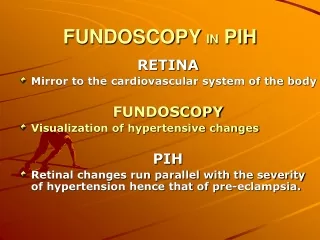
Why do a fundus exam? • To enable detection of the three most common causes of blindness early enough to prevent blindness: 1. Diabetic retinopathy (age group: 20-5o years) 2. Glaucoma (age group: 50-70 years) 3. Age related macular degeneration (70+ years) These are all detectable by examination of the posterior pole of the eye by using a direct ophthalmoscope. Vision-saving treatments are available for these disorders if detected and referred to ophthalmologists early.
ANATOMICAL RELATIONSHIPS • Find the disc!
Direct Ophthalmoscopes Standard Welch Allyn Welch Allyn Panoptic
Tips for successful viewing • Stabilization: • Room lights dimmed • Patient seated, removes eyeglasses; observer standing with eyes at same • level. Observer may leave eyeglasses on • Do not remove contact lenses • Observer at same eye level, standing, with hand on chair or shoulder • Patient should fixate straight ahead • Use your right hand and right eye to view patient’s right eye • Use your left hand and left eye to view patient’s left eye • Grasp handle near the top • Technique: • Center red reflex in view beginning about 10 inches from face • Maintain 15-20 degree temporal angle from direct frontal approach • Rapidly close distance to about 0.5 inch or less from eye (standard scope) • maintaining red reflex centrally in view; with Panoptic place collapsible • tube on face • If disc not in view after 5-10 seconds of viewing, give yourself • and patient a 10 second break • Repeat if necessary until disc is in view; focus if necessary
Find the disc! Using the Panoptic Using the standard Welch-Allyn head
The View White circle: Standard direct ophthalmoscope view Large grey circle: Panoptic view
What am I supposed to do? Have a plan: 1. Examine the disc for: color size sharpness of margins size and shape of cup pattern of disc vessels 2. Look at vessels and their pattern 3. Look at the macular area for: pigmentary changes hemorrhages exudates cotton wool spots 4. Briefly examine all four quadrants Normal disc Normal Macula
Examination of the optic nerve Normal optic nerve Papilledema Note blurring of margin, swollen (choked) appearance tortuosity of vessels, and small hemorrhages
Hypertensive retinopathy Papilledema, papillary hemorrhages, “cotton wool” spots, and narrowed aterioles
Examination of the optic nerve Normal Optic Nerve Glaucomatous Nerve Note size, color, shape, margin, size of cup and lesions Note large cup, nasalization of vessels
The optic nerve in glaucoma Normal Glaucoma
GLAUCOMA Glaucomatous cupping
Examination of the macula Normal macula Background diabetic disease Note absence of landmarks and vascularity Note hemorrhages and exudates
Diabetic retinopathy Advanced background diabetic retinopathy
Examination of the macula Age-related macular degeneration Normal macula Note hemorrhage and pigment changes Fixation marker to locate foveola
Age-related macular degeneration (ARMD) Atrophic ARMD: loss of Retinal pigment epithelium Drusen: early disease