Download
1 / 46
480 likes | 825 Views
Reproductive Endocrinology Tests. Basics Male Tests Female Tests.

E N D
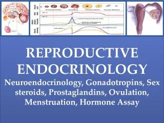
Reproductive Endocrinology Tests Basics Male Tests Female Tests
Abbreviations: AHA, anterior hypothalamic area; AR, arcuate nucleus; DMN, dorsomedial nucleus; MB, mammillary body; ME, median eminence; MN, medial nucleus; OC, optic chiasm; PHN, posterior hypothalamic nucleus; POA, preoptic area; PVN, paraventricular nucleus; SCN, suprachiasmatic nucleus; SO, supraoptic nucleus; VMN, ventromedial nucleus.
Semen Analysis • the volume of semen- 2-6 milliliters • the semen consistency (thickness), • sperm concentration- 40 million spermatozoa per ejaculate • total number of sperm, • sperm motility More than 60% of the sperm show normal forward movement • the number of normal and not normal (defective) sperm- More than 70% N • coagulation and liquefaction - 20-30 minutes after collection • fructose (a sugar in semen), • pH - 7.2-8.0 • the number of immature sperm, and • the number of white blood cells (cells that indicate infection).- No white blood cells or bacteria are detected
The development of a primordial follicle to a preovulatory follicle takes in excess of 120 days. • After it has become a preantral follicle (0.2mm diameter) it takes about 65 days to develop into a preovulatory follicle. • Cohorts of follicles continually develop but only one is ‘selected’ and becomes the dominant follicle. All others undergo atresia.
1 Follicle-Stimulating Hormone • 2 Estrogens • 3 Luteinizing Hormone • 4 Progesterone
Pregnancy Related Diabetes • The prevalence of gestational diabetes mellitus ranges from 1 to 14%. • Screening for gestational diabetes mellitus is strongly advocated at 25-28 weeks of gestation. • This enables early intervention which results in significant improvements in both fetal and maternal outcomes.
Diabetes • The diagnosis can be confirmed by further tests of fasting glucose concentration or • A 75 g oral glucose tolerance test. • These patients should be reassessed in the postpartum period for evidence of diabetes. • The woman's glycated hemoglobin (Hb A1c) should be maintained in the normal range or as near normal as possible to ensure optimal fetal outcome. Normal HbA1c values less than 5%
Pre-eclampsia • Pre-eclampsia occurs typically in the third trimester and affects 4-8% of pregnancies. • It constitutes a triad of: • pregnancy-associated hypertension (that is, there is no pre-existing hypertension), • marked proteinuria (greater than 300 mg daily) and • pathological edema
Pregnancy routine checks • Dipstick Urinary testing for protein, is performed at each antenatal visit together with • Blood Pressure measurement and • careful examination for edema. • Other findings include: rises in serum uric acid (which can antedate the onset of hypertension), urea and creatinine. • Low hemoglobin and platelet concentrations are informative if the patient is suspected to have the severe form of pre-eclampsia - haemolysis-elevated liver enzymes-low platelets (HELLP).
Fetal Health Monitoring • Ultra Sound most commonly used • Alpha fetoprotein • Alpha fetoprotein is a fetal protein arising from the yolk sac and fetal liver. It can be detected in increasing concentrations in maternal serum until 32 weeks of normal gestation.
Neural tube defects • Spina bifida and anencephaly • As a marker of neural tube defects maternal serum alpha fetoprotein, ideally, should be measured between 15 and 18 weeks of gestation.
Down's syndrome • It is the result of either partial or total trisomy of chromosome 21 and is a major obstetric concern, particularly in older women. • Important biochemical markers include: • alpha fetoprotein, • HCG, • unconjugated estriol, • pregnancy-associated plasma protein-A, • serum inhibin-A and • free β-HCG.
Nuchal Translucency (NT) • Increased NT refers to a NT measurement of greater than 3 mm.
While not all birth defects can be found before delivery, certain tests can help to find some birth defects. The options for prenatal diagnosis of birth defects and chromosomally abnormal babies include: • maternal serum triple screen • amniocentesis • chorionic villus sampling • Maternal screening for the detection of neural tube defects and fetal trisomy (most commonly Down syndrome) is offered to women under the age of 35.
Triple/Quad Test • The triple test measures three hormones in the maternal serum: • alpha-fetoprotein • HCG • Estriol • Inhibin-A
Other approaches • Analysis of amniotic fluid: • bilirubin concentration - critical for assessing fetal intravascular haemolysis in the presence of Rhesus incompatability. • Fetal DNA: • A major advance in molecular biology has been the possible detection and isolation of fetal DNA in the maternal circulation. • This exciting discovery has opened up new horizons in the 'non-invasive' assessment of fetal-maternal health. • High concentrations of fetal DNA in the maternal circulation have been found in Down's syndrome, pre-eclampsia, invasive placenta and preterm labour. This technique has also allowed for the prenatal non-invasive diagnosis of • Rhesus D genotype, myotonic dystrophy and achondroplasia
Chorionic Villus Sampling • Chorionic villus sampling involves sampling the chorionic villi, tiny parts of the placenta, either through the vagina or through the abdomen under ultrasound guidance. • Chorionic villus sampling can be performed in the first trimester, usually between 10 and 12 weeks, so the results are obtained earlier than amniocentesis. The pregnancy loss rates after chorionic villus sampling are estimated at 1/100. • In addition, it does not evaluate alpha-fetoprotein, and therefore does not give information about neural tube defects.
A total score of 10 indicates an infant in the best possible condition.An infant with a score of 0-3 requires immediate resuscitation. A low score does not necessarily signify fetal hypoxia-acidosis.
BRCA • The risk at age 50 in BRCA1 carriers is about 50%. • So half of all BRCA1 carriers will already have breast cancer by the time they are 50. • In those that haven’t developed it, the risk will gradually go up as they get older, to a maximum of about 80%. So 80 out of every 100 women carrying a BRCA1 gene fault (8 out of 10) will get breast cancer at some point. And • 2 out of 10 won't. • 20% of women who carry BRCA1 mutations will develop breast cancer by age forty, • 51% by age fifty, and • 87% by age sixty.
The normal semen parameters established by the World Health Organization are:
Laboratory evaluation of amenorrhea • CAH -= congenital adrenal hyperplasia; • DHEAS = dehydroepiandrosterone sulfate; • FSH = follicle-stimulating hormone; • HCA = hypothalamic chronic anovulation; PCO = polycystic ovary syndrome; • PRL = prolactin; T = testosterone; TSH = thyroid-stimulating hormone
PREMATURE OVARIAN FAILURE (POF) • Young women who experience loss of regular menses for three or more consecutive months should be evaluated • Failure to initiate pubertal development by age 13 or begin menstruating by age 15 also warrants evaluation • FSH levels > 30 mIU/mL • Serum estradiol ~50 pg/mL • Basal LH levels > FSH levels (suggests functional ovarian follicles) • Look forhypoestrogenism, and hyper gonadotropinism
PMS • Four common patterns of PMS symptomatology in relation to the menstrual cycle. Note that in every case symptoms commence after ovulation.
PRISM Calendar (Prospective Record of the Impact and Severity of Menstrual symptoms)
Hormonal testing for infertile women is recommended when abnormalities in the menstrual cycle or physical examination are identified. • Tests may include, among many other possibilities, analysis of: • Thyroid function, • FSH and LH levels, • Estrogen or Progesterone levels, Prolactin level or • Androgens such as testosterone, DHEA-sulfate or androstenedione.
Third Trimester (28 weeks – delivery) • Urien screen for sugar and protein • Antibody screen (Rh) • Group-B Streptococcus • Gonorrhea, chlamydia, and syphilis • Hemoglobin and platelet count • Fetal Fibronectin (fFN)
Rh Antibody screen for • Rh-negative mother who has an Rh-positive baby. • If negative for Rh antibodies, then an Rh immune globulin injection (also referred to as RhoGAM), may be given to the Rh-negative mother to prevent her from reacting to the baby’s cells
GB Screen (Group B Streptococcus ) 35-37 weeks • DO NOT CONFUSE WITH Group A (Sore throat) • during delivery can spread to infect the uterus, amniotic fluid, urinary tract • baby may become septic (infection in blood), develop pneumonia, suffer hearing or vision loss, or develop varying degrees of physical and learning disabilities.
Fetal Fibronectin • evaluate a pregnant woman’s risk of preterm delivery\ • fFN, a glycoprotein found at the boundary between the amniotic sac and the lining of the uterus • 26 to 34 weeks • High levels indicate an increased risk
PAP Smears • DNA test for HPV is gaining widespread acceptance as an additional cervical cancer screening tool and as a follow-up test to abnormal changes on a Pap smear. It uses the same cervical sample as the Pap smear to detect and confirm the presence of high-risk types of HPV; it tests for the 13 most common types of high-risk HPV but does not distinguish between them
women 30 years or older be offered the HPV DNA test in addition to their Pap smear and pelvic exam. • If the HPV DNA test and Pap smear are negative and the woman does not have an underlying health condition, she may wait three years before having another Pap smear and HPV DNA test.
Typing of the HPV is not usually necessary. • HPV types 6 and 11 typically cause venereal warts, and (along with types 42, 43, and 44) have a low risk of progressing to cancer. • HPV types 16, 18, 31, 33, and 36 have a higher risk of progressing to cancer.
PAP Test (Papanicolaou smear) • American Cancer Society: Annually • frequent sexual partners, is pregnant, or has abnormal vaginal bleeding, pain, sores, discharge, or itching. • three consecutive normal Pap smears
What does the test result mean? Abnormal changes in squamous cells (ASCUS) or glandular cells (AGCUS) for which the cause is undetermined. An ASCUS test result is frequently followed-up with DNA testing to identify the presence of a high-risk infection with Human Papilloma Virus (HPV).
CIN- Cervical Intraepithelial Neoplasm • The classification of CIN is as follows: • CIN 1: Mild dysplasia • CIN 2: Moderate dysplasia • CIN 3: Severe dysplasia to carcinoma in situ