
ENDOMETRIOSIS
E N D
Presentation Transcript
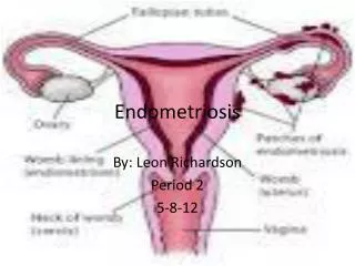
DEFINITIONThe presence of endometrial tissue outside the normal uterus cavity. Endometriosis is Classified Into:Internal type (Adenomyosis): the presence of endometrial tissue in the muscle of the uterus External type: the presence of endometrial tissue outside the uterus or serosal involvement from without
INCIDENCEIncreased in recurrent years due to increased awareness about the disease and availability of laparoscopy as diagnostic tool.
PREDISPOSING FACTORS 1. Age - between 30 - 40 years old 2. Parity - 50 - 700k develop in nullipara3. Estrogen - estrogen may play role in endometriosis 4. Genetic factor - it is common in certain families and in Japanese 5. Socio-economic - it is a disease of rich people 6. Cervical stenosis and RVF 7. Immunological factors - immunological defect (cellular mediated type)
CAUSES OF ENDOMETRIOSIS 1. Retrograde Menstruation (Sampson's theory) Endometriosis is common with cryptomenorrhea against this theory - endometrium, menstruation is non-viable and it can not explain distant endometriosis. 2. SerosalMetaplasia(Meyer's theory) The female genital tract develops from mullerian due
which develop from coelomic epithelium. Endometriosis develop from dormant embryonic cells is differentiated into endometrial cells as a result of inflammatory or hormonal stimuli. Against this theory, it can not explain endometriosis outside pelvis and abdomen, also endometriosis never develop in absence, normal endometriosis.
3. Lymphatic Spread (Halban's Theory) This theory can explain endometriosis, pelvic LN. * Endometriosis of the umbilicus (spread along lymphatic of the urachus) * Endometriosis of the kidney (spread along periureteric lymphatic
4. Diverticular Theory (Cullen's theory) The basal layer of endometriosis grow downwards into myometrium. It is separated from original gland due to uterine contraction. This theory explains adenomyosis, but not endometriosis.
PATHOLOGY OF ENDOMETRIOSIS I. Type (Adenomyosis) * Uterine The uterus symmetrically enlarged but does not exceed 10-12 weeks gestation The wall is thick due to myohyperplasiaIslands of endometrial tissue can be seen scattered throughout the myometrium. They are surrounded with well differentiated stromal cells * Types: - non-functioning type - functioning type (rare) small chocolate cysts are scattered through out the myometrium
II. ExtrauterineA. Pelvic* Ovaries - the most common site to be affected. It is usually bilateral. It takes the form of multiple burnt match head a tarry cyst (chocolate cyst) which is variable in sizes, the wall is thick, granular and surrounded by extensive adhesion
* Serosal surface of the uterus is bickered with multiple small haemorrhagic nodules * Broad ligament, uterosacral and ovarian ligament * Vesico-uterine Pouch and Pouch of Douglas * Fullterm* Frozen pelvis
B. Extra PelvicVulva and vagina in the posterior fornix, RV septum, episiotomy scar Abdominal wall: umbilicus or laparotomy scar Gastro-intestinal tract - caecum, appendix, rectum, recto-sigmoid junction Urinary tract - kidney and uretericPleura and lung
CLINICAL PICTURE SYMPTOMS1. Pain * Dysmenorrhoea - congestive dysmenorrhoea pain occurs 2 - 3 days before the onset of menses due to pelvic congestion, increased after the flow, reaches its maximum in the last day of menses pain is due to pressure if imprisoned blood * Dyspareunia - due to endometriosis of post fornix - RV septum = RVF * Pain during defecation - endometriosis in posterior fornix, RV septum * Acute Abdomen - rupture tarry cyst
2. MenorrhagiaIncreased surface area of endometriumincreased vascularityEndometrial hyperplasia
3. Infertility in 30 - 40% of cases a. Tubal - pelvic adhesions which interfere for with ovum pick up and transport Increased PG content of peritoneal fluid B. Ovarian - AnovultionLPD - due to tutolytic action of PG C. Uterine - production of autoantibodiesunfavourable for implantation d. Peritoneal - macrophages --> phagocytic sperm e. Hyperprolactinaemiaf. Early spontaneous abortion
Examinationgeneral examinationabdominal examination: -ve or endometriosis of umbilicuslocal examination:endometriotic nodule of ovulationendometriotic nodule of recto-vaginal septum uterus symmetrical enlarge or fixed r.v.f.
investigations1. infertility investigation 2. laparoscopy: * confirm the diagnosis * localize and evaluate the extent of lesion * localize and evaluate the extent of adhesion second look laparoscopy at the end of treatment * therapeutic vulva and lysis of adhesion 3. biopsy from skin or vulva lesion 4. curettage in case of menorrhagia5. sigmoidoscopy - git lesion 6. cystoscopy - urinary lesion
Treatment of Endometriosis General Measure 1. get pregnant as early as possible 2. sedative and analgesic 3. iron, vitamin, tonic
Hormonal Treatment I. Induction of PseudomenopauseAim- stop cyclic stimulation of endometrium + atrophy Danazol - 17 alpha ethyl testosterone derivative 1. It acts on the hypothalamus and inhibit release of GnRH2. Inhibit release of pituitary Gn3. Inhibition of ovarian steroid genesis 4. Amenorrhoea5. Dose 200 - 800 mg - 6 months Side effects - acne-hirsutism, weight gain
LH-RH - continuous administration of LH-RH result initial stimulation followed by down regulation (decrease number of receptor) and desensitization (decrease responsiveness) of pituitary gland * Inhibition of release pituitary gland * Inhibition of ovarian steroidogenesis * Amenorrhoea
Dosage - 4 mg microcapsule 1M monthly 3 - 6 months Side effect - expensive, osteoporosis Androgen - testosterone and methyltestosterone Dose: 5 - 10 mg/day 3 - 6 months II. Induction of Pseudo Pregnancy Progesterone - 10 gm/day ---> which increased up to 40 mg/dl Medroxy progesterone acetate
SURGICAL TREATMENT 1. ConservativeResection of endometriotic ---> lysis of adhesion Ventrosuspension of uterus Micro-surgical technique Corticosteroid, antihistamine - postoperative given 2. Laser (X-ray) - used through the laparotomy or laparoscopies for photocoagulation of endometriotic implant
3. RadicalT AH + SSO + Resection of endometriosis Combined Treatment Hormonal treatment followed with surgery Radiological Treatment It is indicated when the operation is risky due to extensive adhesion or endometriosis of recto vaginal septum It is due to deep x-ray therapy or packing of the uterine cavity with radium. It is rarely done.
Chronic Pelvic pain and Endometriosis"Symptoms that depress the doctor". One of these was entitled "Too much pain". Many studies have confirmed that people complaining of chronic pain are more likely to have a neurotic type of personality. Pain has often been found to be a sign of psychological illness and there is no doubt that most patients with chronic pain who present to hospital show evidence of psychological disturbance.
Pain is caused by a condition which has proved resistant to attempts at a cure, the doctor's concern may be increased by feelings of failure and inadequacy. Such is the case with advanced malignant disease. Chronic pelvic pain is a common complaint amongst women in their reproductive years. In addition investigation of pelvic pain was the commonest indication for laparoscopy.
Severe pain is not necessarily caused by organic disease whilst conversely less troublesome pain may be the harbinger of serious pathology. Pain may of course be related to a nongynaecological problem. Table 15.1 Gynaecological Causes of Chronic Pelvic Pain 1. Cyclical pain: DysmenorrhoeaMittelschmerz2. Chronic pelvic inflammatory disease 3. Endometriosis 4. Neoplasia: Benign - fibroids - ovarian cyst Malignant disease of the genital tract
5. Pelvic venous congestion I pelvic pain syndrome (PPS) 6. Polycystic ovarian syndrome 7. Residual ovary syndrome 8. Uterovaginalprolapse
Table 15.2 Non-gynaecological causes of chronic pelvic pain Intestine:DiverculitisInflammatory bowel disease Irritable bowel syndrome Malignancy Subacute intestinal obstruction
Urinary tract: Calculus Chronic retention Infection Malignancy Musculo-skeletal: Lumbo-sacral osteoarthritis Prolapsed intervertebral disc Spondylolisthesis
INNERVATIONGynaecological pain may be somatic, from the vulva, perineum and lower vagina, and transmitted via the pudendal nerves (S2, 3 and 4) or visceral from the uterus, fallopian tubes, ovaries and visceral peritoneum supplied by the autonomic nervous system. Visceral and somatic pain perceptions are different. The viscera are insensitive to thermal and tactile. Referred pain results from irritation of the overlying peritoneum, and is perceived in dermatomes supplied by the same nerve root.
Stimuli that produce pain include the following: 1. Distension and contraction of a hollow organ 2. Rapid stretching of the capsule of a solid organ 3. Chemical irritation of the parietal peritoneum 4. Tissue ischaemia5. "Neuritis", secondary to inflammatory, neoplastic or fibrotic processes in adjacent organs
Characteristics of the Pain: 1. Mode of onset 2. Duration 3. Site 4. Radiation 5. Relationship to menstrual cycle and previous pregnancy 6. Intensity: this can be best assessed by instructing the patient in the use of a visual analogue scale
EXAMINATION The formal examination should cover the following points: 1. General examination, specifically looking for signs of malignancy such as lymphadenopathy, anaemia and swelling of the lower limbs. 2. Abdominal palpation for masses, ascites and tenderness. Vaginal examination: 3. Speculum examination Bimanual palpation 4. Rectal examination, to exclude malignant disease 5. Examination of lumbo-sacral spine and hip joints
GYNAECOLOGICAL CONDITIONS Cyclical Pain Dysmenorrhoeais a common complaint, although only in 5%. In the majority of cases there is no underlying pathology although congestive dysmenorrhoea is said to be associated with such conditions as endometriosis or adenomyosis. Ovulation Pain (Mittelschmerz) mid-cycle is of acute onset and is a sharp lower abdominal pain by several hours of dull aching in the pelvis. Caused by rupture of the ovarian follicle at ovulation, the onset of pain corresponds to the peak LH levels 24 hours prior to ovulation perifollicular smooth muscle, mediated through prostaglandin.
PELVIC INFLAMMATORY DISEASE (PID) Clinical signs and symptoms are frequently misleading however. In women who have a combination of lower abdominal pain, vaginal discharge and pelvic tenderness only 600k will be found to have pelvic infection at laparoscopy.
ENDOMETRIOSIS The pain of endometriosis is variable in presentation and there is a poor correlation with the laparoscopic findings. Severe pain may be associated with minimal disease, whilst the reverse is also true. The commonest symptoms are dysmenorrhoea, dyspareunia and persistent dull ache in the pelvis, although severe acute pain may be caused by the rupture of an endometriotic cyst.
NEOPLASIA OF THE GENITAL TRACTBenignLarge fibroids may cause a persistent dull aching pain in the pelvis, whilst an ovarian cyst may produce recurrent episodes of sharp pain secondary to torsion of the ovarian pedicle.
MalignantAlthough pain is not a leading feature of malignant disease of the cervix and body of the uterus, lower abdominal and pelvic pain is the commonest presenting symptom in advanced ovarian cancer.
Pelvic Pain Syndrome (PPS)Dull aching pain with occasional severe acute attacks more commonly present in the right iliac fossa, but sometimes moving from one side to the other.
Polycystic Ovarian Syndrome (PCOS) It has been postulated that the excess oestrogen seen in PCOS causes dilatation of the pelvic veins (Reginald et al 1989), as oestrogen has been shown to inhibit the contraction of smooth muscle in the walls of human veins.
Residual Ovary Syndrome Symptoms from ovaries left at the time of hysterectomy. Chronic pelvic pain and dyspareunia are the presenting symptoms in approximately 75% of cases.
UterovaginalProlapseA dull aching pain or dragging sensation in the pelvis in association with a "lump" in the vagina.
NON-GYNAECOLOGICAL CONDITIONS GastrointestinalDiverticular disease is common in people over the age of 60 years and approximately half the patients have chronic or intermittent lower abdominal pain. Constipation, or alternating
diarrhoea and constipation with passage of pebbly stools may accompany the pain or occur independently. The irritable bowel syndrome (TBS) is a common complaint of women presenting with pelvic pain to a Gynaecology clinic.
Renal Tract Causes of pain arising from the urinary tract are infection and the presence of calculi. Chronic infection can result in persistent pelvic pain, whilst chronic interstitial cystitis usually occurs in middle-aged women and causes severe supra-pubic pain.
Ureteric and Bladder Calculi May cause recurrent episodes of lower abdominal and pelvic pain. Skeletal Causes Pelvic pain are often polysymptomatic and may well complain of back-ache. Low back-ache of musculo-skeletal origin radiates most commonly to the lower limbs and not to the abdomen or pelvis. Hip pain may sometimes be mistaken for pain arising in the lower back and characteristically is most severe in the groins but may also be felt in the buttocks.
INVESTIGATIONS LaparoscopyLaparoscopy is by far the most informative and is an essential part of the investigation in all cases of pelvic pain.
ImagingUltrasound Is a very informative investigation which has the added advantage of being non-invasive. The dimension of uterus and ovaries tend to be larger than normal in cases of PPS whilst the diameter of pelvic veins can be measured using the Doppler mode.
X-Rays1. Plain x-ray of lumbo-sacral spine and hip joints 2. Intravenous urogram3. Barium enema 4. Transuterine pelvic venographyComputerized Tomography and Magnetic Resonance Imaging Although these techniques have no place in assessing the majority of women with chronic pelvic pain they are invaluable in assessing the spread of malignant disease.
OTHER INVESTIGATIONS 1. Examination of mid-stream urine specimen 2. Full blood count may reveal an iron deficiency which although commonly caused by menorrhagia, may alert to the possibility of a colonic malignancy.