Download
1 / 17
200 likes | 999 Views
AP Biology Classification Project Porifera,Cnidarians,Rotifera. By: Amin Syed and Marwa Saleh. PORIFERA- THE SPONGES. Structure :.

E N D
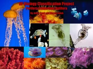
AP Biology Classification Project Porifera,Cnidarians,Rotifera By: Amin Syed and Marwa Saleh
PORIFERA- THE SPONGES Structure: The outer layer of a sponge is called the epidermis layer. They are made up of epithelial cells. Porocytes regulate water flow and they are located around pores. Some portions of the layers contract (some pores close) when they are touched or exposed to chemical stimuli. The Internal cavity (spongocoel) has specialized flagellated cells called Choanocytes (collar cells). The lining of the choanocytes creates an inner layer (gastrodermis). These cells line the entire body cavity. Sponges lack symmetry but very few small sponges are radially symmetrical Poriferas are multicellular which means that their cells are specialized for different functions of the body Sponges are sessile (they do not move)and they attach themselves to something solid, but larval sponges are free-swimming. Sponges also consist of mesohyl between body cavity and the outer wall Mesohyl are gelatinous, protein-rich matrix. That’s where amoeboid cells are located. Amoebocytes secrete hard mineral needles called spicules and tough protein fibers called spongin. Both of these structures strengthen and protect the sponge, provide the support to keep the pores open. Osculum which is a large pore where water and chemicals leave. Sharon. (2011). The porifera (sponges). Retrieved from http://sharon-taxonomy2010-p2.wikispaces.com/Porifera
PORIFERA- THE SPONGES Diversity: Mode of nutrition: • Sponges are classified in the kingdom animalia. • The only phyla is Porifera but there are four different classes. They are Calcarea (chalky sponges with calcium carbonate), Demospongiae (they include glass sponges & Venus flower basket with spicules), Hexactinellida (they include horny & bath sponges with only spongin or spongin/silica spicules) ,and Sclerospongiae (they are coral sponges. They have spongin, silica, and calcium carbonate spicules). • The subkingdom is Parazoa which means that they lack tissue and organs. They also lack nervous system. • There are approximately 5150 species • Sponges are filter feeders. They remove food (plankton) from water which is brought through the pores (Ostia) • Then the flagella pulls in bacteria or algae which then stick to the collars of choanocyte. They are then passed to archaeocytes who digest food. • The amoebocytes travel in the mesohyl and pick up the food from choanocytes and distribute to other parts of the sponge. Because of the amoebocytes, then finally water and food that are not needed leave through the osculum which is like a large hole on top of the sponge. • They are heterotrophic consumers, they ingest food meaning they digest internally.
Reproduction: PORIFERA- THE SPONGES • Sponges can produce asexually and sexually. • They produce asexually by external buds. They either break off to form new sponges or they stay attached to form sponge colonies. • They also have specialized internal buds called Gemmules. Gemmules consists of food-filled amoebocytes by a coat with spicules, they break their coat when there are no harsh conditions and form new sponges. Sponges are hermaphrodites (they produce both eggs and sperm) but they exchange sperm. • Mesohyl Is the location where sexual reproduction occurs. Male gametes are released into water by the sponge and then it goes through the pores of the neighboring sponges. Spermatozoa sticks to the collar of choanocytes, the choanocytes then lose their collar and the collar turns into a specialized cell similar to amoebocytes cells that carry spermatozoa to the egg. The fertilized egg becomes the blastula (free-swimming larvae) and is released into water. They settle and then are transformed into adult sponges. • Sponges can also reproduce by regeneration, where missing body parts are regrown. No genetic modification techniques used because sponges are not commonly used organisms. Sharon. (2011). The porifera (sponges). Retrieved from http://sharon-taxonomy2010-p2.wikispaces.com/Porifera
PORIFERA- THE SPONGES Ecological significance: • Some sponges may be used to find out new pharmaceutical compounds that can be used on humans. • Because they are natural filters, sponges can remove pollutants and carbon dioxide. • They serve as protective houses for mollusks and small fishes. Small fishes who live inside the sponge can take advantage of the food supply that is circulating in the water. • They have commensal relationship with shrimps because shrimp live inside them and grow so large that its hard for them to leave through the osculum. • Sponges are pathological towards human like bacteria or fungi can be. So they are harmless towards humans. • They can be used as products for cleaning • Several sponge species produce compounds that show great potential to be used as a drug to cure malaria, tuberculosis and other infectious diseases. • Also, you can get a skin rash from sponges. Due to their defense mechanism of releasing toxins. Uniqueness: They have a natural filter feeding system. Simplest mutlicellular organism. They possess choanocytes and special flagellated cells whose beating drives water through the body cavity.
Examples: PORIFERA- THE SPONGES • Tube Sponge: are very common and can be found in the coral reefs. they are different because of they are long tube-shaped and range in color from purple to blue, gray, and gray-green. They are one of the few reef invertebrates that are blue In color. • Vase Sponge: common species found in the Caribbean. They have a large bell shape with deep central cavity. grow up to 2 feet wide and 3 feet high. ranges in color from purple to red and brown, and most of them are attached to sandy bottoms. • Yellow Sponge: are commonly found through out the Pacific coastal waters. Are found growing in small colonies, and ranges in color from orange to bright yellow. They encrust rocks on reefs. • Red Tree Sponge: are common throughout the Caribbean Sea. They grow up to height of about 8 inches. They are very easy to keep and can live easily in home aquarium environment. Also, they require moderate water flow and dim light.
Structure: Rotifera- Wheel bearer • The foot ends in a toe which contains cement gland- useful because it attaches itself to objects in water and obtain food when it wants to. • They are transparent and are covered with a cuticle covering. This tells us that they are closely related to roundworms and arthropods. • The food is chopped by trophi (jaws) which is located in the behind the mouth in the pharynx (throat). • Head is the corona or crown of cilia which draws water into mouth and sifts for food. They also use cilia to move themselves – some of them can walk with head and foot. • They are bilaterally symmetrical. There are four basic regions: head, neck, body, and foot. • They have a body cavity which is lined by mesoderm. • They have specialized organ systems and a complete digestive tract which includes mouth and anus. • For their nervous system, their nerves extend through out their body. They have five eyes with one or many photoreceptors. • They have small brains and contain one ore two antennae.
Rotifera- Wheel bearer Mode of nutrition: Diversity: • They are very tiny- 200 to 500 micrometers long • Microscopic organisms. • They are the most common microorganisms collected from ponds and small lakes. • Their habitats are freshwater, marine, or damp soil. • They are divided into three classes: Monogononta, Bdelloidea, and Seisonidea. • The food particles are very small so it can pass through their mouths. Examples include unicellular algae and phytoplankton. • They complete their digestive tract through gastrovascular cavity. They use the alimentary canal (a digestive tube with a separate mouth and anus). • The crown of cilia draws water into the mouth and pharynx contains trophi or (jaws) that grind up food, mostly microorganisms suspended in water. • For circulation, movement of the rotifer body distributes the fluid through out the body which then circulates the nutrients. It has an open circulatory system. • There is no specific organ for gas exchange so the only way is through diffusion.
Rotifera- Wheel bearer Reproduction: • reproduce asexuallly by parthenogensis, in which females produce more females from unfertilized eggs • other species might produce two types of eggs that develope by parthenogenesis. One type forms females,other type develops when conditions deteriorate and become simplified males that can not even feed themselves • males only survive long enough to fertilize eggs, which form resistant zygotes that can survive when a pond dries up • zygotes break dormancy and develop into a new female generation
Rotifera- Wheel bearer Ecological Significance: Uniqueness: • Important ecolofical role as suspension feeders on phytoplankton and bacteria • Trophic Link-food source for larval fishes • Sewage treatment • Ephemeral Waters-Ability to survive dessication • Only reproduce asexually • seudocoelomates • fluid in their pseducoelom serves as a hydrostatic skeleton • smaller than protists, but more anatomically complex than flatworms
Rotifera- Wheel bearer Examples: • Philodina roseola–This class of rotiferas do not have a cuticular covering, males have never been observed, and the females appear to be obligatorally parthenogenic. They can survive extremes of temperature and desiccation for years. As these were the first rotifers to be described, they were given the common name of "Wheeled animacules". Class Bdelloidea. • Rotifer neptunis-is 1 mm long when fully extended, but can retract its body like pushing in a telescope until it is a third of this length. The females produce live young. Class Bdelloidea. • Hydratina senta- This is the marine Class; they are relatively large and live in the gills of crustaceans.Class Seisonidea. • Keratella quadrata-This class of rotifera contains the largest number of species. There are both freshwater and marine species. Sexual reproduction has been observed, although males are absent for most of the year, and much smaller than females. Class Monogonata
Cnidarians-Jellyfish,Hydra,Corals and Sea Anemones Structure: • Cnidarians have two body forms, Polyp and Medusa. These two phases alternate in the life cycles of the cnidarians. But a number of the cnidarian species exist only as polyps for example the Corals and Sea anemones. • Both forms have two layers, the epidermis (the outer layer) and the gastrodermis ( the inner layer that has the digestive tissues. These two layers are separated by the mesoglea, a geleatinous material that holds the muscles for most cnidarians. • Polyps are cylindrical and are usually found attached to a substrate on the floor of ocean. The mouth faces away from the substrate the polyp is attached to, therefore often faces upward with its tentacles pointing upward. Polyps build up a chitinous or calcareous external or internal skeleton or polyp or both. • Medusae are free-floating and umbrella shaped. Their mouths point downwards surrounded by their venomous tentacles. • The tentacles and sometimes the body surface bear cnidocyte cells, which secrete Nematocyst, which is a barbed or venomous coiled thread that can be projected in self-defense or to capture prey.
Cnidarians-Jellyfish,Hydra,Corals and Sea Anemones Diversity: Mode of Nutrition: • Cnidarians are classified in the animialia kingdom. • Cnidarians have three main classes: Anthozoa (corals) ,Hydrozoa (hydras) and Scyphozoa (jelly fish). • Anthozoa-can be either solitary or colonial. Common forms include corals and sea-anemones. They differ from other Cnidarians in that they have no medusoid stage. • Hydrozoa-have calcified skeletons of aragonite and calcite. • Scyphozoa-only occur in marine environments. And the majority of species in this class are jellyfish. • There are approximately 10,000 specimens of cnidarians • Cnidarians vary in diet due to the variety of different species. • Their diet ranges from, phytoplankton to small fish. • For specifically jellyfish, Nematocysts fire on contact; after paralyzing its prey, it pushes the food into its mouth with its tentacles • Food is pushed into the central cavity where gland cells release enzymes in order to digest food. • Other cells of the endoderm will then absorb the digested nutrients. Indigestible material will then be eliminated out of the mouth.
Cnidarians-Jellyfish,Hydra,Corals and Sea Anemones Reproduction: • Both the polyp and medusae forms consist of diploid individual. • Polyps reproduce asexually, by simply budding with the neighboring polyps. When doing this they can either produce another polyp or a medusae. • Cnidarians exhibit open-water fertilization • Medusae reproduce sexually. The male organisms release sperm into the water and it is collected by the female, who then releases the fertilized eggs that become free swimming larvae called planulae.
Cnidarians-Jellyfish,Hydra,Corals and Sea Anemones Ecological Significance: Uniqueness: • Coral reefs are a shelter for many marine animals. • Coral reefs provide habitat for many fish and food for many other animals that produce valuable shells, pearls, and jewelry. • Medical research of chemicals produced by many Cnidarians, contributed in the creation of new antibiotics and anticancer chemicals • They were among the first multicellular life to evolve on earth. • They have been here for at least 650 million years. • Their most obvious unique feature is their highly specialized stinging cells.
Cnidarians-Jellyfish,Hydra,Corals and Sea Anemones Examples: • CLASS HYDROZOA-Members of this class include the Hydra, Obelia, and Gonionemus. Most members of this class have both the polyp and medusa stages yet, Hydra exists only in the polyp form. • CLASS SCYPHOZOA -Animals in this class are entirely marine. The polyp stage is reduced or absent. This class mainly includes jellyfish species. • CLASS ANTHOZOA - the term anthozoa means "flowering animals“. They exist in the sessile polyp stage only; no medusa stage is present. This class includes the sea anemone and sea coral. • CLASS CUBOZOA-have a square shape when viewed from above. They also have four evenly spaced out tentacles or bunches of tentacles.
References • Fautin, Daphne G. and Sandra L. Romano. 1997. Cnidaria. Sea anemones, corals, jellyfish, sea pens, hydra. Version 24 April 1997. http://tolweb.org/Cnidaria/2461/1997.04.24 in The Tree of Life Web Project, http://tolweb.org/ • Morris, Matthew May 15 2003. Phylum Cnidaria. http://www.angelfire.com/mo2/animals1/phylum/jellyfish.html • Ward, Paul. 2005-2012. Cnidaria – Cnidarians. http://www.darwinsgalapagos.com/animals/cnidaria_jellyfish_coral_sea_anemone.htm/.http://www.darwinsgalapagos.com • McGraw-Hill companies.(2002). The NoncoelomateAnimals. In Raven/Johnson, Biology(pp.884-889). New York: McGraw-Hill Publisher. • McDarby Michael. (2001-2011). An online introduction to the biology of animals and plants [Web log message]. Retrieved from http://faculty.fmcc.suny.edu/mcdarby/animals&plantsbook/animals/02-Sponges&Cnidaria.htm • bumblebee.org. (1997-2012). Retrieved from http://www.bumblebee.org/invertebrates/Porifera.htm • Myers, P. 2001. "Porifera" (On-line), Animal Diversity Web. Accessed February 20, 2012 at http://animaldiversity.ummz.umich.edu/site/accounts/information/Porifera.html • Sea sponges. (2012). Retrieved from http://www.allthesea.com/Sea-Sponge.html • Baqai , A. (2000, June 08). Retrieved from http://www.ucmp.berkeley.edu/phyla/rotifera/rotifera.html • Sharon. (2011). The porifera (sponges). Retrieved from http://sharon-taxonomy2010-p2.wikispaces.com/Porifera • bumblebee.org. (1997-2012). Retrieved from http://www.bumblebee.org/invertebrates/ROTIFERA.htm • Integr. Comp. Biol. (2002) 42 (3): 660-667. doi: 10.1093/icb/42.3.660 . http://icb.oxfordjournals.org/content/42/3/660.full