Download
1 / 72
730 likes | 787 Views
Neuroendocrine tumors (NETs) are complex to classify due to organ specificity, increasing incidence, and varying clinical presentations. This article delves into the definition, epidemiology, clinical symptoms, pathological classification, and the role of pathologists in diagnosing and managing NETs. The text provides insights into the origin of NETs, their epidemiological aspects, common sites of manifestation, and the challenges of staging and grading. Additionally, it discusses the importance of immunohistochemistry in diagnosis and the multidisciplinary approach required for effective management.

E N D
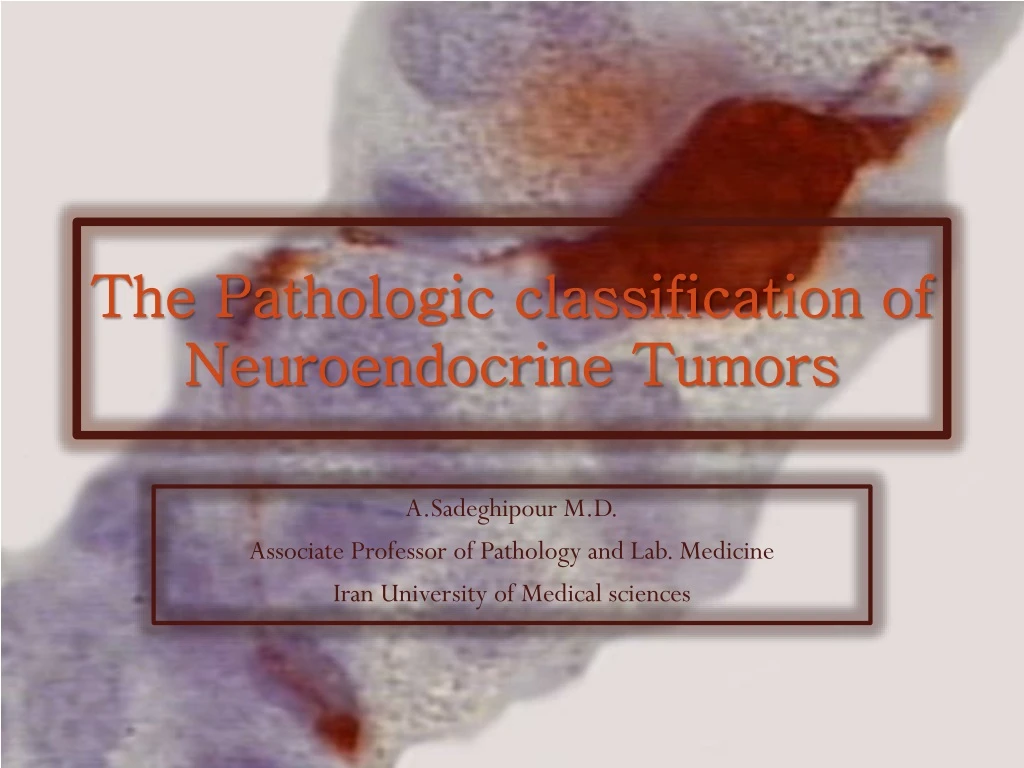
The Pathologic classification of Neuroendocrine Tumors A.Sadeghipour M.D. Associate Professor of Pathology and Lab. Medicine Iran University of Medical sciences
50%OF THE NEUROENDOCRINE TUMORSARE DIAGNOSED WHEN IN ADVANCED STAGES
Complexities of Organ Specific Classification of NETs Could Lead to Confusion inDiagnosis and Management
Neuroendocrine TumorsOverview of the Presentation • Definition and history • Epidemiological aspects • Clinical presentation • Paraclinical diagnosis • Immunohistochemistry • Pathological classification, staging and grading • Role of Pathologist in multidisciplinary approach to diagnosis and management
Neuroendocrine TumorsDefinition • Neoplasms originating from the neuroendocrine cell system characterized by cytoplasmicneurosecretory granules Enterochromaffin Cell
Embryology • Embryologic data have now refuted the neural crest origin of most neuroendocrine cells.
NETs • NETs arise from cells which produce and secrete hormones • Most NETs are slow growing and malignant, with metastatic potential • Common sites of origin are: • GI tract • Lungs • Pancreas
600 6.00 Incidence of all malignant neoplasms 5.25 Incidence of neuroendocrine tumors 500 5.00 400 4.00 Incidence of all malignant neoplasms per 100,000 Incidence of NETs per 100,000 300 3.00 2.00 200 1.00 100 0 0 76 78 80 82 84 86 88 90 92 94 96 98 00 02 04 74 Year Incidence of Neuroendocrine Tumors Is Increasing • The incidence and prevalence of NETs has increased approximately 500% over the past 30 years (better diagnosis?) Source: US SEER database. Adapted with permission from Yao JC, et al. J Clin Oncol. 2008 26:3063-3072.
NETs Are the Second Most Prevalent Type of GI Malignancy in the US 1,200, 000 1,100, 000 2x more prevalent than pancreatic cancer 2x more prevalent than pancreatic cancer 100, 000 [ ] [ 0 Colorectal1 GEP-NET2 Stomach1 Pancreas1 Esophagus1 Hepatobiliary1 Prevalence in SEER Database 1. National Cancer Institute. SEER Cancer Statistics Review, 1975-2004. http://seer.cancer.gov/csr/1975_2004. 2. Modlin IM, Lye KD, Kidd M. Cancer. 2003;97(4):934-959.
NET Neuroendocrinetumours (NETs) are derived from cells that have the unique ability to synthesise, store and secrete a variety of metabolically active substances, peptides and amines, which can cause distinct clinical syndromes.
Tumours regarded as NETs on the basis of the immunohistochemical expression of markers of NE differentiation Endocrine-Related Cancer (2010) 17 R173–R193
Neuroendocrine TumorsClinical Presentations • Asymptomatic cases (50%) • Initial clinical symptoms are often non-specific and variable • Carcinoid syndrome (symptoms mistaken with other more common problems) • Most NETs are non-secretory and their clinical presentation before metastasis is non-specific
GI NETsClinical Presentation • Clinical presentation of GI NETs varies widely • Often discovered incidentally • Symptoms due to: • Mechanical bulk • Fibrosis • Secretion of various hormones • Serotonin is most common substance secreted from GI NET • Serotonin release can cause carcinoid syndrome Serotonin-producing GI NET (ileum)
Hypotension Flushing Rapid heart beat Heart disease Wheezing Diarrhea GI NETsCarcinoid Syndrome • Symptoms of carcinoid syndrome include: Carcinoid syndrome is associated with metastatic disease
Natural History of Neuroendocrine Tumours Estimated time to diagnosis: 5 to 7 years Death * Diarrhea Vague abdominal symptoms * Flushing Metastases Primary tumour growth 1 2 3 4 5 6 7 8 9 Time, years *Symptoms of carcinoid syndrome Vinik A, et al. Pancreas. 2009 Nov;38(8):876-89 19
Nonspecific Symptoms Are Commonto Multiple Diagnoses Menopause Functional Bowel Disease Symptoms • Sweating • Flushing • Diarrhea • Intermittent abdominal pain •Hypoglycemia• Confusion • Bronchoconstriction • Dyspepsia •GI bleeding • Cardiac disease Food Allergy Irritable Bowel Syndrome Neurosis Alcoholism NET Peptic Ulcer Asthma Anxiety Thyrotoxicosis 20 Aggarwal G, et al. Cleve Clin J Med. 2008;75(12):849-855.
Spectrum of paraneoplastichumoral syndromes secondary to NETs
Functional NETs: Secrete active substances , peptides and amine • Non-Functional NETs: Mass effect
Neuroendocrine Tumors Pathological classificationStaging/Grading
ClassificationTraditional Method • NETs have been traditionally classified as foregut, midgut, or hindgut, depending on site of origin • Replaced by new tumor-based classification method developed by World Health Organization (WHO)
Classification World Health Organization Method • WHO classification defines NETs by degree of tumor differentiation, with specific clinicopathological features • The WHO classification system is based on the following criteria: • Biological behavior (malignancy) • Metastases • Ki-67 index • Angioinvasion • Tumor size • Histological differentiation • Hormonal syndrome
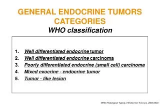
WHO Classification 2000 • The nomenclature used in this classification is considers the following factors: • Histomorphology (degree of differentiation) • Presence / absence of local invasion / metastasis (Stage) • Proliferation index (grade)
Well differentiated endocrine tumors - low grade malignancy Well differentiated endocrine carcinomas Poorly differentiated endocrine carcinomas - small cell carcinomas Mixed exocrine and endocrine carcinomas Tumor-like lesions WHO Classification 2000
Neuroendocrine tumor grade 2 RadioGraphics March 2002 vol. 22 no. 2 351-365
Small cell neuroendocrine carcinoma grade 3 Cesar A. Moran etal. Am J Clin Pathology, 2009; 131: 206-221.
Large cell neuroendocrine carcinoma Grade 3 Cesar A. Moran etal. Am J Clin Pathology, 2009; 131: 206-221.
WHO Classification 2010: NeuroendocrineNeoplasms of the Digestive System • Working principles • “Neuroendocrine” defines the peptide hormoneproducing tumors and share neural-endocrine markers • “Neuroendocrine neoplasm” includes well- and poorly differentiated tumors • Premise: All neuroendocrineneoplasmshave a malignant potential • This premise has an influence on the incidence data because NENs that were regarded as benign and not considered in the incidence data (eg, SEERS data) now have to be included
WHO Classification 2010: NeuroendocrineNeoplasms of the Digestive System • Main criteria determining the malignant potential –Tumor histopathology o Well differentiated o Poorly differentiated –Proliferative activity G1, G2, G3 –Site, size, infiltration/invasion, metastasis (TNM) o Esophagus, stomach, duodenum, ileum, appendix, colorectum,pancreas
NeuroendocrineNeoplasms: NENs of the Gastroenteropancreatic (GEP) System Bosman FT, et al. WHO Classification of Tumours of the Digestive System. Lyon, France: IARC Press; 2010.
NeuroendocrineNeoplasms: NENs of the Gastroenteropancreatic (GEP) System Bosman FT, et al. WHO Classification of Tumours of the Digestive System. Lyon, France: IARC Press; 2010.
Systems of Nomenclature for Neuroendocrine Tumors (Pancreas & Volume 39, Number 6, August 2010) The grade of the tumor MUST be included in the pathology report, along with a reference to the specific grading system being used. Unqualified terms such as neuroendocrine tumor or neuroendocrine carcinoma without reference to grade do not provide adequate pathology information.
Diagnostic Standards for NENs • Mandatory: • Histopathology―well or poorly differentiated • Expression of neuroendocrine markers synaptophysin and chromogranin A • Proliferative activity: G1–G3 • Stage: pTNM (ENETs 2007 and UICC 2009)
Neuroendocrine TumorsIHC for Diagnosis • Cytosolic (NSE, PGP 9.5) • Related to secretory granules (chromogrnins) • Related to synaptic vesicles (synaptophysin, VMAT) • Intermediate filaments (NF, High molecular weight CK) • Adhesion molecules (N-CAM)