Download
1 / 50
590 likes | 1.37k Views
Acute Coronary Syndromes. Jeffrey Smith, M.D., M.P.H. George Washington University Medical Center Jim Holliman, M.D. Pennsylvania State University. Epidemiology. Ischemic Heart Disease (IHD) is the leading cause of morbidity and mortality worldwide

E N D
Acute Coronary Syndromes Jeffrey Smith, M.D., M.P.H. George Washington University Medical Center Jim Holliman, M.D. Pennsylvania State University
Epidemiology • Ischemic Heart Disease (IHD) is the leading cause of morbidity and mortality worldwide • Over the next 10 years, IHD will become the leading cause of death and disability in most developing countries • IHD is the leading cause of death in USA ; 1 million annually • 50 % of all cardiovascular deaths occur in women • Despite educational efforts, patients with Acute Myocardial Infarction (AMI) delay an average of 2 to 6 hours before seeking health care
Diagnosis remains a challenge • Of the 1.25 million AMI’s in the USA yearly, over 80 % present to the Emergency Department (ED) • The history, exam and ECG can be nondiagnostic • Past studies : 5 to 8% of AMI’s are sent home • Recent studies : 2 to 5% of AMI’s are sent home • Mortality doubles when AMI’s are sent home
Acute Coronary Syndromes (ACS) • Maintain high clinical suspicion • Early recognition • Early risk stratification Optimize therapy
4 Areas of Delay • Patient seeking care • Early triage with ECG • Delay in clinical decision to treat • Treatment • Aspirin, oxygen, beta-blocker, Unfractionated heparin or Low Molecular Weight Heparin • Thrombolytics (door to drug in under 60 minutes) • Intervention (door to cath lab in under 2.2 hours)
ACS : Clinical Syndromes • Angina • Class I (severe exertion) to IV (with any activity or at rest) • Fixed stenotic lesion ; stable precipitating and relieving factors • Unstable Angina Pectoris (USAP) • Increasing frequency, transition to a higher class, pain at rest • Variant (Prinzmetal’s) angina • AMI • 2 of 3 W.H.O. criteria : • clinical history or setting, ECG changes, positive cardiac markers • Non-ST Elevation (NSTE) ACS versus ST Elevation Myocardial Infarction (STEMI) • Various reperfusion and adjunctive therapies are based on the presence of ST segment elevation in the appropriate clinical setting
Pathophysiology of ACS • Endothelial damage occurs by plaque disruption, irregular luminal lesions or shear injury • Platelet aggregation is followed by thrombus formation • Ischemia causing local mediator release and vasospasm • Reperfusion injury • In USAP spontaneous thrombolysis occurs rapidly. • Persistence of occlusive thrombus for 60 to 90 minutes before spontaneous lysis results in a non-STE AMI. • Transmural AMI occurs when the thrombus occlusion lasts for 2 to 3 hours.
History & Physical……and Atypical Presentations • History is essential … but not diagnostic of ACS • only 20 to 30 % report crushing or pressure pain • Beware of atypical presentations • Up to 20 % of ACS present with pain in other areas (arm, back, abdomen) • No pain in 25 % of diabetics • Burning pain in 15 % of USAP or AMI (may get relief with antacids) • Right sided radiation in up to 30 % • Chest wall tenderness in 15 % of AMI’s • Anginal equivalent symptoms in 30 % of AMI’s • Dyspnea, nausea, diaphoresis, back pain, jaw pain • Altered mental status (especially if > 85 y/o) • Mortality 3-fold greater (50 % vs 18 %) • High risk groups : young, elderly, diabetic • Physical Exam most helpful in excluding noncardiac causes
Differential Diagnosis • Causes of chest pain • Most are noncardiac and not serious • Acute coronary syndrome • Pulmonary embolism • Aortic dissection • Pericarditis / tamponade • Pneumothorax / tension pneumothorax • Pulmonary parenchymal process : pneumonia, inflammatory process, benign and malignant processes
Diagnostic Studies • ECG • Serial ECG’s • Cardiac enzymes • Risk Stratify for other studies based on history, ECG, and above studies
The Electrocardiogram • Advantages: • select appropriate therapy • determine response to treatment • determine inpatient disposition • predict complications / death
The Electrocardiogram • Beware that the 12-lead ECG is diagnostic of transmural AMI in only 25 to 50 % of cases • Non-specific ST / T-wave abnormalities • 6 % of transmural AMI’s • 30 % of NSTE-AMI • 20 % of cases of unstable angina • Completely normal in 3 to 4 % of transmural AMIs • Serial ECGs may increase sensitivity by 10 to 20 %
The Electrocardiogram • Nondiagnostic ECG seen in 50 % • Nonspecific ST-Twave (NSSTTW) changes • Less than 1 mm ST depression • Blunted, flattened or biphasic T waves • Low risk of AMI (4 %) yet 20 % risk of USAP • If good story & NSSTTW changes : ADMIT • LVH, LBBB, VPR may mask signs of acute ischemia
The Electrocardiogram • Low-risk criteria (14 % AMI, 0.6 % Cardiac events, 0 % mortality) • Normal ECG • NSSTTW changes • Unchanged ECG • High-risk criteria (42 % AMI, 14 % Cardiac events, 10 % mortality) • Ischemic ST segment or T wave changes • LVH, LBBB, VPR
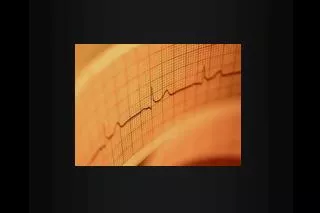
Q : ECG manifestation of STE- AMI • Increase R wave voltage • Hyperacute T wave evolves to STE over the next 30 minutes • Pathologic Q waves : • Commonly develop 24 hours after infarct May not occur in up to 50 % of patients with an acute myocardial infarction. • Serial ECG if persistent pain, atypical symptoms or change in VS. (or ST segment trend monitoring)
Differential diagnosis of this ECG tracing • Acute myocardial infarction • Hyperkalemia • Subarachnoid hemorrhage • Benign Early Repolarization (BER) • LVH • Hypertrophic cardiomyopathy • Acute pericarditis
Cardiac Markers • Cardiac Panel • Myoglobin • CPK • CPK-MB • Troponin (T and I) • Rapid bedside assays available
Cardiac Enzymes • Myogobin • Increased levels within 2 to 4 hours • Peaks at 6 to18 hours • Returns to baseline by 24 hours • Elevated levels also found in skeletal muscle injury (e.g. rhabdomyolysis)
Cardiac Enzymes • CPK • Increased levels within 6 to 8 hours • Peaks at 12 to 24 hours • Returns to baseline by 36 to 48 hours • Elevated levels also found in cases of skeletal muscle injury (e.g. rhabdomyolysis) • CPK-MB • Similar time course as for total CPK • More specific for cardiac muscle • Most specific if elevated and 5 % of total CPK
Cardiac Enzymes • Troponin T and I • Increased levels within 6 to 8 hours • Peaks at 18 to 24 hours • Troponin T remains elevated for 5 to7 days • Troponin I remains elevated for up to 10 to 14 days • More specific for cardiac muscle (troponin I is most specific) • Mild elevations seen in up to 1/3 of cases of unstable angina ( “microinfarcts”) • Prognostic value for cardiovascular complications • By 12 hours after onset of symptoms, CPK-MB and Troponin I have a 100 % sensitivity for detecting AMI
Additional studies for diagnosing AICS • 2-D echocardiogram • Useful with a nondiagnostic ECG or difficult ECG’s • Sensitivity 85 % ; specificity 50 % • Can’t distinguish ischemia, AMI or previous infarction • Less sensitive for nontransmural. • Radionuclide scanning : thalium 201, technetium-99 • Decreased uptake in ischemic tissues • Normal perfusion and stress nuclide scans carry very low risk • ETT • The more abnormal the baseline ECG, the less useful the test • Useful in low and intermediate risk groups stratification and discharge
Risk Stratification TIMI risk Score Age > 65 Documented coronary stenosis > 50 % Three or more risk factors Two or more anginal equivalents within 24 hours ST segment changes Increased cardiac biomarkers High risk features : Elevated cardiac biomarkers Diabetes TIMI score > 5 Refractory symptoms despite medical management Elevated C reactive protein Relative risk features : Prior CABG Prior MI LV dysfunction CHF Management
STEMI : Aspirin 162 mg LMWH or UFH Clopidogrel 300 mg load Nitroglycerin Morphine Metaprolol Lytic therapy If PCI : Abciximab Revascularization (PCA / stent) NSTE-ACS If No high-risk features : Aspirin LMWH Clopidogrel If Positive high risk features : Add IIb-IIIa GP inhibitor AHA 2002 Guidelines
Other Treatments • Ticlopidine • Inhibits tranformation of IIB-IIIa glycoprotein receptor • Good for USAP : takes 8 to 10 days to reach maximal benefit. • Recommend to load with 300 mg initially (unless CABG anticipated) • LMW heparin • Better than unfractionated heparin for USAP • Better inactivation of Factor Xa • Less nonspecific binding • Longer half-life, predictable response • Less bleeding complications
Glycoprotein IIb-IIIa receptor inhibitors • Monoclonal antibodies that more effectively inhibit platelet function than aspirin. • Completely inhibit platelet adhesion • Proven additive benefit in patients undergoing percutaneous intracoronary vascular procedures (abciximab). • Increasing evidence that these agents may be helpful in ischemic episodes not responsive to other agents. • ACC / AHA 2002 guidelines for management of NSTE ACS : • Level 2A recommendation for patients without intended early cath / PCI
Management Sequence for ACS Pt arrives to ED Targeted history Physical assessment, V.S., EKG ASA 160 mg PO Oxygen, IV assess Cardiac monitoring & send labs
Management of Chest Pain (CP) If patient continues with CP and not hypotensive : Nitro 0.4 mg SL q 5 min X 3 prn Morphine 2 to 4 mg IV q 5 min prn Beta Blocker (Metoprolol 5 mg IV q 5 min x 3) LMWH, Clopidogrel CEU Protocol USAP, NSTE-ACS Admit Thrombolytics IIb-IIIa GP inhibitor, PCI
Thrombolytics • Reduce short-term mortality by 18 to 25 % • Overall : • All agents are effective and should be combined with antithrombin agents • Larger the infarct, the greater the mortality reduction with thrombolytics • Accelerated t-PA results in better flow at 90 minutes and 15 % reduction in 30 day mortality • TNK (mutant form of t-PA) is as effective as accelerated t-PA and single bolus dosing. TNK is 14 x more fibrin specific, 80 x more resistant to PAI-1
Thrombolytics : eligibility criteria • Consistent history and physical exam • STE > 1 mm in two or more contiguous limb leads or > 2 mm in two or more contiguous precordial leads (not ST depression : not ischemic syndromes !) • New LBBB • Therapeutic window is 12 hours from symptom onset.
Contraindications (CI) to Thrombolytics • Absolute CI : • Prolonged CPR • Major surgery or trauma within the last 10 days • Significant coagulopathy • Diabetic hemorrhagic retinopathy • Left heart thrombus, SBE, pericarditis • Oral anticoagulant use • Septic thrombophlebitis • Previous CVA • Relative CI : • > 12 hours after onset of symptoms • Age > 75 ??? • Poorly controlled hypertension (> 180 to 200 / 110 to 120)
Additional reperfusion strategies • PCI (percutaneous coronary interventions) • Lower risk of intracranial bleeding • Higher reperfusion rates • Rapid evaluation of patients not eligible for thrombolytics • Better risk stratification, earlier discharge • Early identification of surgical candidates • Prior CABG patients (grafts thrombose, larger burden of clot) • Superior to thrombolytics if intervention within 90 minutes • Recommended for patients in shock if PCI in 90 to 120 minutes. • Rescue PCI if poor response to thrombolytics • Grab bag of various strategies with no clear conclusions : i.e. lose dose thrombolytics prior to immediate PCI (facilitated PCI), glycoprotein inhibitor plus thrombolytics, etc. : Long term benefit yet to be proven. • Radioactive implants
Who to transfer ? • Transfer to a tertiary care facility with interventional and cardiac surgery capabilities is indicated : • CI to thrombolytics and may benefit from PCI, CABG • Persistent hemodynamic instability or ventricular arrhythmias • Ongoing ischemia following infarction or thrombolytics • Patients SHOULD receive the initial standard ED therapy including thrombolytics.
Final Thoughts • Rapid history, rapid ECG with interpretation, rapid treatment • Maintain a high clinical suspicion : 2 to 10 % of patients with CP and AMI are released from ED • Young, atypical presentations, nondiagnostic ECG’s • Single ECG and single set of enzymes can give a false sense of security • ED cocktail : ASA, MSO4, β-blocker, clopidogrel, LMWH, IIb-IIIa agents • Target goal of thrombolytics within 30 minutes for STEMI • Strongly advocate for PCI when appropriate
A Comprehensive Approach : Goals of the ED Cardiac Evaluation Unit (CEU) • Early Systematic Evaluation of All Chest Pain Patients • Identification of all reperfusion candidates • Reduction of delays in initiation of reperfusion therapy • Improved diagnosis and treatment of other dangerous pathology • Early (primary) risk stratification
ED Evaluation of Chest Pain • 5,000,000 visits / year in U.S. • 5 % of all ED visits • 5 to 15 Chest Pain patients / day per ED • Lengthy Differential Diagnosis
Identifying the Problem : Need for a More Aggressive Approach to Acute MI : • “Time is Myocardium” • Dramatic Mortality Reduction in STEMI with Early Reperfusion • Thrombolytics • Immediate PTCA / stenting • Increasing number of other treatments available for ACS • Require early risk assessment
Team Members • Cardiologist : specialist, algorithm • ED Physician : team leader • algorithm, educate & motivate staff • Senior triage nurse : team facilitator • algorithm, educate & motivate staff • Clinical technician : algorithm, educate & motivate staff • Patient Service Coordinator : algorithm • Research coordinator : data collection
CEU Goals (Continued) • Accelerated Rule-out of MI among low risk patients • Reduce LOS • Improve cost-effectiveness • Improve patient satisfaction • Eliminate “missed MI” • Secondary Risk stratification • Comprehensive Diagnosis and Treatment Center • Education and Community Outreach • Multidisciplinary approach
Implementation :Goal 1 : Early Diagnosis of MI • ALL chest pain patients receive immediate, aggressive evaluation • Vital signs • Cardiac monitor • Intravenous “lifeline” placed • Oxygen and/or pulse oxymetry • ECG performed under standing orders within 10 minutes of arrival • ECG handed directly to physician for interpretation
Goal 1 : Early Diagnosis of MI • Early focused history and physical exam • Blood drawn upon arrival for initial myocardial markers (Cardiac Panel) • CK-MB • Troponin • Myoglobin • “Reperfusion Checklist”
Goal 2 : Evaluation of Low Risk Patients • “Zero tolerance” for missed MI • MI can not be excluded by history, exam alone • Hospitalization for “Rule-Out MI” expensive, inefficient • Several alternative approaches now exist
Goal 2 : Evaluation of Low Risk Patients Initial Risk Stratification within 1 to 3 hours : • ST segment elevation MI : • Thrombolytics or cath lab • High Probability for ACS and / or High Risk : • Ischemic ECG, positive marker, etc. • ICU vs in-patient unit, anti-ischemic therapy, ? cath • Low Risk for ACS : • No high risk clinical features • Non-diagnostic ECG • Initial myocardial markers negative • Hold for further workup or go to CEU • Negligible Risk : • Atypical history, no risk factors, normal ECG and markers, other cause identified • Discharge home
Goal 2 : Evaluation of Low Risk Patients • Continuous observation on cardiac monitor • 12 to 24 hours • Accelerated “rule-out” of MI • Serial ECGs • Serial Myocardial Marker Measurements • 0, 3, 6, 9 hours • CK-MB, troponin • MI definitively excluded within 12 hours
Goal 3 : Secondary Risk Stratification • MI rule-out does not exclude other ACS (Unstable Angina) • Further evaluation often needed prior to discharge • Stress testing • Screening test for CAD • Accurate predictor of short and long term risk of death, adverse events • Reduces need for repeated evaluations
Stress Testing • Standard (Bruce protocol) exercise (treadmill) stress testing • Stress echocardiography • Radionucleotide scanning • Thallium • Sestamibi • Dual isotope • Pharmacologic stress testing
Low Risk Chest Pain Evaluation Protocol 1 hour 3 hours 6 hours 9 hours Initial Risk Stratification History Physical Exam ECG Myoglobin CK-MB Troponin ECG ECG ECG (+) Admit Diagnostic Stress Testing Low Risk Patient All Negative (-) Home Continuous Monitoring CK-MB CK-MB CK-MB, cTn
Impact of CEU Approach :Low Risk Chest Pain Patients Problems : “Unnecessary” admission of low risk patients, “Missed” MI’s Measure : length of stay, % MI’s discharged Interventions : accelerated rule-out protocol Impact : Missed MI’s reduced from 4.5 to 0.4 %* Length of stay reduced ~ 2.5 days to 1 day * Graff et al. Am J Cardiol 1997; 80 : 563-8