Download
1 / 17
170 likes | 277 Views
Measurement of Spleen Stiffness to Evaluate Portal Hypertension and the Presence of Esophageal Varices in Patients With HCV-Related Cirrhosis GASTROENTEROLOGY Dr Azad bakht.

E N D
Measurement of Spleen Stiffness to Evaluate Portal Hypertension and thePresence of Esophageal Varices in Patients With HCV-Related CirrhosisGASTROENTEROLOGYDr Azad bakht
Portal hypertension (PH) is a frequent complication of cirrhosis, contributing to the development of ascites, esophageal varices (EV), and hepatic encephalopathy.
The hepatic vein pressure gradient (HVPG) is the standard used to determine the degree of portal hypertension (PH) and an important prognostic factor for patients with cirrhosis.
. However, HVPG can only be accurately determined at specialized centers; noninvasive methods are needed to predict HVPG values and the presence of EV.
However, the performance of HVPG is limited to highly specialized centers and requires extensive experience and therefore is not used routinely.
splenomegaly in cirrhosis is characterized by enlargement and hyperactivation of the splenic lymphoid tissue, as well as increased angiogenesis and fibrogenesis, in addition to passive congestion due to PH. spleen stiffness (SS), assessed by magnetic resonance elastography.
SS values were obtained using the FibroScan with the same probe used to perform LS after at least 6 hours of fasting and under US assistance. In the absence of guidelines for the measurement of SS by FibroScan, the same guidelines for the measurement of LS were applied.
the aim of this study was to assess the relationship between SS measured by TE (FibroScan; Echosens, Paris, France) and PH in terms of HVPG together with the possibility of predicting the presence of EV according to SS in a cohort of 100 consecutive patients with hepatitis C virus (HCV)-related cirrhosis without clinically evident complications. The study was performed in a tertiary center: the Department of Clinical Medicine of the University of Bologna (Italy).
Each patient was studied according to a 3-day protocol as follows: day 1 included clinical and biochemical evaluation, complete abdominal US, and measurement of LS and SS by TE; day 2 included measurement of HVPG; and day 3 included upper digestive endoscopy.
patients with a splenicparenchymal thickness of <4 cm under the probe were excluded. Other exclusion criteria were the presence of ascites, severe obesity, and the absence of an intercostal space sufficiently wide for the use of the FibroScan probe.
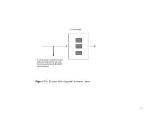
. Figure 1. Flow chart of the studied patients
A first interesting observation of this study is the high diagnostic accuracy of SS, in terms of AUROC, for both the diagnosis of EV and the definition of the degree of PH (HVPG ≥10 or ≥12 mm Hg). Particularly concerning the diagnosis of EV, an SS cutoff value <41.3 shows an LR− of 0.029 and thus seems able to accurately rule out the presence of varices and support a possible screening strategy, reserving esophagogastroduodenoscopy only for patients with an SS >41.3.
even more interesting is the possibility to assess noninvasively HVPG by measurement of SS and LS. In fact, with a simple linear model including both variables, it seems possible to obtain an accurate estimate of HVPG. Predictive equation: HVPG = −4.44 + 0.241 • LS + 0.226 • SS.
SS reflects the changes in splanchnichemodynamics leading to the formation of EV better than LS and any other noninvasive parameter investigated in the present study.