Download
1 / 50
500 likes | 717 Views
Chemical Power for Microscopic Robots in Capillaries. Tad Hogg Institute for Molecular Manufacturing. with Robert A. Freitas Jr. preprint: http://arxiv.org/abs/0906.5022. Sensors for Medicine and Science (SMSI) implantable glucose monitor. Given Imaging’s PillCam photos inside GI tract.

E N D
Chemical Power for Microscopic Robots in Capillaries Tad Hogg Institute for Molecular Manufacturing with Robert A. Freitas Jr. preprint: http://arxiv.org/abs/0906.5022
Sensors for Medicine and Science (SMSI) implantable glucose monitor Given Imaging’s PillCam photos inside GI tract Aarhus University targeting drugs to cells microscopic robots for medicine • extending today’s implanted devices • to much smaller sizes • enhancing today’s in vivo nanoparticles • with computation & communication
challenges formicroscopic robots • fabricate • control • power • identify applications
power • short operation: • fuel inside robots • extended operation: • power from environment, e.g., • acoustics (ultrasound) • chemical fuels from environment see R. Freitas, Nanomedicine Vol. I, chap. 6, 1999
example: glucose + oxygen C6H12O6 + 6O2 6CO2 + 6H2O energy released: 4x10-18 Joule per reaction e.g,: react a million O2/second = about a pico watt
How much power can glucose + oxygen provide?
Is power generation safe for nearby tissue?
method • pick medical application context • identify key constraints on power • evaluate quantitatively
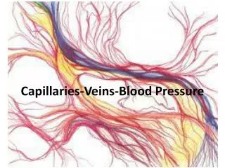
application: robots in bloodstream • useful for diagnosis and treatment • devices pass near cells throughout body • examples • passive motion with fluid • active motion • self-assembled groups on vessel wall • for long term monitoring
glucose and oxygen in blood • blood plasma • glucose concentration ~100x that of oxygen • hence: oxygen is rate-limiting chemical • most oxygen carried in red cells • bound to hemoglobin • ~100x higher concentration than in plasma
chemical power in capillaries • modeling chemical transport • results • robot power • effect on surrounding tissue
modeling challenges • complex geometry • distorting cells • kinetics of oxygen release from cells
geometry • simplify to axial symmetric • vessel, robots and tissue
Krogh cylinder model tissue cylinder capillary venous end flow arterial end A. Krogh, J. of Physiology 52:409 (1919)
numerical model domain 100μm capillary radius 4 μm 40 μm model a portion of ~1mm capillary length
robots inside vessel 10μm 8μm static robot scenario: robots attached to wall fluid moves past the robots example robot group: each robot about 1μm3 20 robots in each ring 10 rings group of 200 robots
modeling challenges • complex geometry • distorting cells • kinetics of oxygen release from cells
cells distort to go through capillaries “single file” cell maintains volume and surface area as it distorts typical values: surface area 135 μm2 volume 90 μm3
approximation for cell distortion group of robots vessel wall plasma gap direction of flow vessel wall distorted cells based on Secomb et al., American J. of Physiology: Heart and Circulatory Physiology 281:H629 (2001) instead of modeling individual cells, use “smeared out” average fluid with mix of cells and plasma
modeling challenges • complex geometry • distorting cells • kinetics of oxygen release from cells
oxygen transport oxygen diffuses out of cell into plasma from plasma into tissue O2 cell O2 flow diffusion: from higher to lower concentrations hence cell still has substantial oxygen at end of vessel
cell oxygen release & robots • robots have high power densities • larger concentration gradients than tissue • cells may pass before releasing oxygen • model must include kinetics
cell oxygen release kinetics • depends on • concentration in surrounding plasma • amount of oxygen bound within cell • approximate model Clark et al., Biophysical Journal 47:171 (1985)
numerical model • fluid flow • oxygen transport • from cells • diffusion in plasma and tissue • power generation • in robots and tissue • Michaelis-Menten kinetics • heating • conduction and convection
chemical power in capillaries • modeling chemical transport • results • robot power • effect on surrounding tissue
scenarios • low demand • resting tissue • slow flow • avg. speed 0.2mm/s • high demand • active tissue • fast flow • avg. speed 1mm/s typical situation for nanomedicine
oxygen transport to robots and tissue
section through vessel and tissue tissue vessel wall flow streamlines of fluid flow tissue distance (μm)
no robots (1022 molecule/m3 ) flow distance (μm)
10-micron group (1022 molecule/m3 ) flow distance (μm)
no robots (1022 molecule/m3 ) flow distance (μm)
10-micron group (1022 molecule/m3 ) flow distance (μm)
power comparisons • robots ~10s of pW • cells use 10-1000pW • cell size ~10mm • i.e., ~103 x robot volume • person at rest uses ~100 watts
What can a picowatt do? • compute: ~105 logic operations/sec • near-term molecular electronics • 103 kT/operation • communicate: ~104 bits/sec over 100mm • with ultrasound • move: ~1mm/sec through water • overcoming viscous drag see R. Freitas, Nanomedicine Vol. I, chap. 6, 1999 www.nanomedicine.com
effects of robots • less oxygen for tissues • local heating • waste products • forces on vessel wall • …
oxygen for tissue • robots • compete with tissue for oxygen • block diffusion out of vessel • is this a problem for nearby tissue?
(1022 molecule/m3 ) examine tissue power next to vessel wall flow distance (μm)
power drop ~ 10% power in tissue next to vessel flow compare no robots 10-micron group power decrease much less than oxygen concentration decrease due to Michaelis-Menten kinetics parameters in tissue (oxygen must get very low to cause significant power decrease)
heating • robots have high power density ~107 watt/m3 • tissue cells ~104 watt/m3 • possible significant local heating
temperature increase by robots high demand 10-4 degrees C flow
heating not significant • about 10-4 degree C • for a group of 100s of robots • heating would be significant if: • much larger groups • many groups in nearby capillaries
summary • ~10s of pW per robot on capillary walls • for group of ~100s of robots • oxygen: mainly from passing cells • small reduction in tissue power • especially for low demand scenario (resting tissue) • insignificant local heating • in spite of high power density
additional biology questions effects on robots and surrounding tissue
local effects • white blood cells • block oxygen transport as they pass • additional forces on robots • other functions of blood • e.g., immune response, clotting • response of cells in vessel wall to • forces from robots clinging to wall • blocked chemical transport
long-range effects • lower cell oxygen downstream of robots • response to partially blocked vessel • increase pressure? • time scale of response • systemic effects of many robot groups
further info • T. Hogg, Designing Microscopic Robots for Medical Diagnosis and Treatment, • Nanotechnology Perceptions3:63-73 (2007) • preprint: http://arxiv.org/abs/0906.5022 • R. Freitas Jr.,www.nanomedicine.com