AdrenoLeukoDystrophy
AdrenoLeukoDystrophy. Blong Yang Molecular Genetics Fall 2004. University of Wisconsin – Eau Claire Fall 2004. Adrenoleukodystrophy (ALD). What is Adrenoleukodystrophy:


AdrenoLeukoDystrophy
E N D
Presentation Transcript
AdrenoLeukoDystrophy Blong Yang Molecular Genetics Fall 2004 University of Wisconsin – Eau Claire Fall 2004
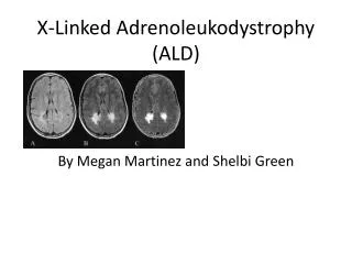
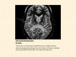
Adrenoleukodystrophy (ALD) What is Adrenoleukodystrophy: • X-linked genetic disease that is a peroxisomal disorder with impaired beta-oxidation of saturated Very Long Chain Fatty Acids (VLCFA’s). - First recognized in 1923 – Schindler’s Disease Siemerling and Creutzfeldt - 1970 – Dr. Michael Blaw first coined the term Adrenoleukodystrophy Adreno = Referring to adrenal glands Leuko = Referring to white matter of the brain Dystrophy = Referring to imperfect growth/development • Caused by the X-ALD gene that encodes the ALD protein - Peroxisomal membrane protein - Member of the ATP binding cassette transporter protein superfamily. - Demonstrated using mouse model for X-linked ALD - Function that contributes to ALD is still unknown Lombard-Platet et al. Heinzer et al. University of Wisconsin – Eau Claire Fall 2004
X-ALD Phenotypes Frequency of ALD in human population: • Affects 1/20,000 males There are 2 Major disease Phenotypes: • Childhood onset ALD • Most severe form of ALD • Affects boys between the age of 4 – 6 years • Accounts for ~35% of ALD patients • Involves inflammatory demyelination of nerve cells • Leads to a vegetable state • Death within a few years Lombard-Platet et al. University of Wisconsin – Eau Claire Fall 2004
Born May 1993 and appears as a normal healthy boy. Age 3, difficulties Potty training. Age 7, has difficulties at school, fighting, constantly getting lost, difficulty learning material and keeping attention in class. MRI diagnose Alex with ALD Age 8, Alex can no longer walk. Loses sight, muscle coordination, ability to speak and swallow. Currently, Alex is still alive and undergoing muscle therapy and on muscle relaxants, baclofen and Clonidine. Meet Alex………. University of Wisconsin – Eau Claire Fall 2004
X-ALD Phenotypes • Adult onset ALD or Adrenomyeloneuropathy • Occurrence at a mean age of 27.6 +/- 8.7 years. • Most common, accounting for ~ 65% of ALD patients • Progresses slowly • Adrenal impairment • Involves mainly long tracts of the spinal cord and peripheral nerves • No involvement of the Brain • Little or No inflammation • Learning disabilities, perceptual problems, attention deficit disorder, short and long-term memory loss, impaired vision, coordination or gait, and various personality and behavioral changes. • Addison Disease • Impaired Adrenal Function • Adrenocortical Insufficiency with no detectable neurological involvement • “Bronzing” of the skin Lombard-Platet et al. University of Wisconsin – Eau Claire Fall 2004
Location and Cloning of X-ALD Gene • First cloned in 1993 • Cloned by Mosser et al. • Method: Positional Cloning • Approach: • Hypothesize that the location of X-ALD gene must be Xq28, where the red/green color pigment gene (R/GPG) resides. On the basis of the high incidence of colour vision anomalies in AMN and ALD patients, the ALD and R/GPG must be close. • Identify a patient (Patient O) with blue-monochromatic colour vision with a rearrangement located 5’ of Re-Colour pigment (RCP) gene, including two deletions separated by a large (>100 kb) inversion. • Only the RPC gene is found on the first deletion (88 kb). This deletion was not found in 81 other ALD patients, therefore they postulated that the inverted repeat or the second deletion were candidate loci for the ALD gene. Mosser et al. University of Wisconsin – Eau Claire Fall 2004
Location and Cloning of X-ALD Gene • BP4-BP2 and BP3-BP1 shows breakpoint 4 and 2 and of points 3 and 1. • Dotted line represents the breakpoint junctions cloned. • Use a probe 4 kb proximal to Fr15 deleted in Patient O (15) to obtain clones from a Xq28 cosmid library. • 3 overlapping clones were obtained spanning 90 kb (cosQc11H12, cosQc8F3 and cosQc14A11) • In parallel, an Xba1 junction fragment (X-8) was cloned corresponding to BP4-BP2 in patient O. A Map of the ALD gene Mosser et al. University of Wisconsin – Eau Claire Fall 2004
Location and Cloning of X-ALD Gene Generating Probes • Restriction map of X-8 showed a 1.8 kb Xbal-EcoR1 (X2) fragment. It contained a 1.6 kb segment on the BP4 side and is within the Fr15 contig. • Use Taq1 to generate probes from digestion of cosQc11h12. • Probes generated are Ta25 (2.1 kb), Ta4 (3.6 kb), Ta1 (1.0 kb), Ta18 (0.85 kb), Ta13a (935 bp), Ta13b (252 bp). • Probes detected deletions in 5 patients. Patient O, B, R, L and Family Ma. Mosser et al. University of Wisconsin – Eau Claire Fall 2004
Figure d shows segregation of an abnormal junction fragment detected by probe X2 in family. Here, you can see a 14.4 abnormal fragment was detected in an affected male as well as heterozygous females. Figure e shows 3 brothers whose DNA is digested with HindIII and probed with X2. The affected brother, filled square shows an abnormal 22 kb fragment. The brother affected shows the same 22 kb fragment. Unaffected brother doesn’t show 22 kb fragment. Location and Cloning of X-ALD Gene Southern Blot Mosser et al. University of Wisconsin – Eau Claire Fall 2004
Sequencing the ALD gene • Use probes TA25, TA18, TA13a and TA13b to probe for transcripts in several tissues. • Discovered a 4.2 kb transcript. • The sequences for these probes determined (Using a computer program based on a multiple sensor-neural network approach.) • Sequencing revealed a large 700 bp putative protein coding region for TA25 and between 300 – 400 bp regions for TA18, TA13b and TA13a • Discovered that the deduced amino-acid sequence from these sequences showed significant sequence identity with collinearly positioned regions of human and rat 70 K peroxisomal membrane protein (PMP). Mosser et al. University of Wisconsin – Eau Claire Fall 2004
Using Nested - P.C.R reactions with primers from putative exons produced two fragments. Ex13 and Ex3. These fragments where then used to screen a random-primed HeLa cell cDNA library to obtain 6 independent overlapping clones, Producing a 2,751 bp sequence encoding a 745 aa protein.
Protein Homology Alignment of the c-terminal amino-acid sequence of ALD Protein with human and rat 70 K PMP and other members of The ATP-binding protein family. Identical are in black, Conservative substitutions are in gray. The two ATP binding Domains are underlined, the segment highly conserved in ABC proteins are indicated by asterisks. University of Wisconsin – Eau Claire Fall 2004
Conclusions drawn by J. Mosser et al. • The putative location of the ALD gene is in the distal part of Xq28. • The ALD gene does not code for VLCDF – CoA synthetase which was once considered to be the cause of ALD. • The ALD gene codes for a protein that is almost identical to human and rat 70 K Peroxisome membrane protein. • The ALD protein is a member of the ATP – Binding Cassette (ABC) transporter protein, which are involved in the transport of proteins, amino acids, inorganic ions and peptides. • The ALD must either nonfunctional in transport of VLCFA or it might be inhibited from activating VLCFA – CoA synthetase. • It is still unresolved why the brain and adrenal glands are so sensitive to the buildup of VLCFA. Mosser et al. Heinzer et al. University of Wisconsin – Eau Claire Fall 2004
Map of the ALD gene The X-ALD gene is approximately 20 kb long and consists of 10 exons. Mutations to the ALD gene that have been discovered can be found at http://www.x-ald.nl/ . To date, there are 703 mutations in their Database.
Mouse model for X-linked ALD NO!!! YES!!! • - J.F. Lu et al. In 1997 • Use mouse model to show that the ALD gene leads to the accumulation • of VLCFA. • -Method – Disruption of the ALD gene with Neo-R gene. University of Wisconsin – Eau Claire Fall 2004
Disruption of the X-ALD mouse gene Depiction of X-ALD gene exons 1-4. Gene targeting of the X-ALD locus used a replacement- Type vector covering between Dashed lines. Homologous recombination Causes disruption in the X-ALD Gene in ES cells. Grown on selective medium containing Gangcyclovir. Select surviving cells. Use XbaI to digest and run On southern blot and Probe For sequences. Wildtype ES has 7 kb sequence, Mutant has 8 kb sequence. University of Wisconsin – Eau Claire Fall 2004 J.F. Lu et al.
Disruption of the X-ALD mouse gene Using neo-r cassette-specific primers (e or b) in combination with external primers (a or f) ES cells 1-3 generated a 4 kb PCR product but no results for normal ES cell. Using internal primers (d or c) in combination with external (a or f), wildtype produced a 3 kb PCR product while mutant ES 1-3 cells produced the 4 kb (included the neo-r gene). University of Wisconsin – Eau Claire Fall 2004 J.F. Lu et al.
Production of Chimeras and transmittance of X-ALD gene • The targeted ES cells are injected into blastocysts derived from C57bl/6 strain and the blastocysts were transferred into pseudo-pregnant CD-1 female mice to generate chimeras. • Female gave birth to 9 chimeras (6 males, 3 females) • Males are crossed to C57BL/6 to transmit X-ALD- allele to progeny giving rise to heterozygous female carrier mice. • Heterozygous female mice crossed with either 129/Sv strain or C57bl/6 strain to generate hemizygous male mice (X-ALD-/ 0). • Among the progeny: • Normal sex ratio (Male:Female = 110:102) indicating hemizygotes are not embryonic lethal. • Out of 110 male mice, 54 were X-ALD-/ 0. University of Wisconsin – Eau Claire Fall 2004 J.F. Lu et al.
There is a great difference in the amount of total VLCFAs in normal and X-ALD mice and humans as well, especially when the FAs contain more than 26 carbon units. University of Wisconsin – Eau Claire Fall 2004 J.F. Lu et al.
Histological Analysis Performed at 3 months. Slide A shows healthy normal adrenocortical cells. Slide B shows an X-ALD-/ 0 mouse adrenocortical cells with clear lipid clefts (indicated by arrows). Slide C shows the lipid cleft admixed with spicular lamellar material at high magnification. (x 15,000) University of Wisconsin – Eau Claire Fall 2004 J.F. Lu et al.
Conclusion of Mouse Study • The X-ALD gene leads to the accumulation of VLCFAs in various tissues including the brain, kidneys, pancreas, liver and heart. But the level of accumulation of each tissue varies. • The oldest mice at the end of the study were 6 months old and did not show any symptoms of adrenal insufficiency or nervous system dysfunction. It is yet too early to conclude that the X-ALD will not have any neurological involvement equivalent to that of human X-ALD. • Biochemically, the X-ALD mouse model represents a valid animal model for X-ALD. Most importantly, X-ALD in both mouse and humans result in elevated tissue levels of saturated VLCFAs, with the most severe abnormalities localized to the brain and adrenal gland. University of Wisconsin – Eau Claire Fall 2004 J.F. Lu et al.
Diagnosis and Treatment of ALD • Diagnosis: • Simple blood tests can be done to analyze the levels of VLCFAs in males. This test is not very accurate for female carries. • Genetic testing is available through a DNA-based blood test. This test is accurate in testing for carriers. The entire testing protocol is available through the United Leukodystrophy Foundation (ULF). • Treatment: • There is no definitive treatment to ALD • Lorenzo’s Oil diet (not FDA approved) • Bone Marrow Transplant • Gene therapy has been developed in animal models, but is currently not available to human patients. University of Wisconsin – Eau Claire Fall 2004 J.F. Lu et al.
One more thing…….. Reviews: “Ebert and Roeper” - Two thumbs up!- University of Wisconsin – Eau Claire Fall 2004
References: • Heinzer, A.K., Kemp, Stephan., Lu, J.F., Watkins, P.A., Smith, K.D. “Mouse Very Long-chain Acyl-CoA Synthetase in X-Linked Adrenoleukodystrophy.” J. Biol. Chem. Vol 277, Issue 32, 28765-28773 (2002) • Heinzer, A.K., Watkins, P.A., Lu, J.F., Kemp, Stephan., Moser, A.B., Li, Y.Y., Mihalik, S., Powers, J.M., Smith, K.D. “A Very Long-chain Acyl-CoA Synthetase Deficient Mouse and Its Relevance to X-linked Adrenoleukodystrophy.” Human Molecular GeneticsVol. 12 No. 10, 1145-1154 (2003) • Lombard-Platet, Gael., Savary, Stephane., Sarde, C.O., Mandel, J.L., Chimini, G. “A Close Relative of the Adrenoleukodystrophy Gene Codes for a Peroxisomal Protein With a Specific Expression Pattern.” Proc. Natl. Acad. Sci. USA 94, 1265-1269 (1996) • Lu, J.F., Lawler, A.M., Watkins, P.A., Powers, J.M., Moser, A.B., Moser, H.W., Smith, K.D. “A Mouse Model for X-linked Adrenoleukodystrophy.” Proc. Natl. Acad. Sci. USA 94, 9366-9371 (1997) • Mosser, J., Douar. A., Sarde, O., Petra, K., Feils, R., Moser, H., Poustka, A.M., Mandel, J.L., Aubourg, P.“Putative X-linked Adrenoleukodystrophy Gene Shares With Unexpected homology with ABC Transporters.” Nature 361, 726-730 (1993) University of Wisconsin – Eau Claire Fall 2004