Download
1 / 34
340 likes | 386 Views
Initial Evaluation of Trauma Patient in Emergency Department and ATLS

E N D
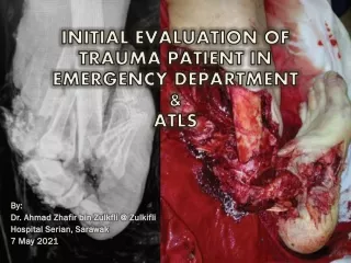
INITIAL EVALUATION OF TRAUMA PATIENT IN EMERGENCY DEPARTMENT & ATLS By: Dr. Ahmad Zhafir bin Zulkfli @ Zulkifli Hospital Serian, Sarawak 7 May 2021
Lethal triad • The understanding that all the process started at scene and by the time pt arrive ETD - acidosis sets in etc. • Current trend - if need blood product - early blood product, not recommended to give too much crystalloids.
Scenario • It was a rainy Friday at 4.45 pm in the evening. You are sitting at the MO table at ETD, settled patients admission and just referred a patient to SGH for CT brain - CVA TRO ICB (golden hour). Your other colleagues came to ETD just lingering around awaiting to punch their attendance card. • Suddenly, your ETD phone rang. Your overzealous assistant medical officer answered. He then informed to you: “ Boss, MACC just called. We got one male patient about 20-30 years old, Honda City skidded at Pan Borneo BalaiRingin, victim was inside car, conscious but pale and very weak, unable to move, holding his abdomen. EMS KK BalaiRingin responded and currently bringing the patient to our ETD, ETA 15 minutes.” • Damn, what to do?? (silently praying that patient will arrive after 5 pm)
Pre arrival preparation • Patient profile • Mechanism • Vital signs • Apparent injuries • Early notification enables emergency department staff to do the following: • Notify additional personnel • Assure resources are available • Prepare for anticipated procedures • Prepare for blood transfusion Watch out for ineffective care plans and mismanagement (teamwork): Communication breakdown Failure in situation awareness Staffing and workload distribution Unresolved conflicts between staffs
Patient arrived at 4.55pm (five minutes early..) • You are getting passover from the paramedic while your staffs transferring the patient. You quickly recalled MIST: • Mechanism (and time) of injury • Injuries found and suspected • Symptoms and Signs • Treatment initiated • It was Mr. William Shakespeare, 24 years old Iban male from Kampung Antayan, car driver with seat belt on, his car skidded around 4.15pm. After received call from public, EMS team arrived at 4.30pm. He was removed from the driver seat and patient was screaming pain at his abdomen. EMS team suspected intrabdominal injury. He was put on cervical collar and spinal board and brought to ETD Serian. Blood pressure at ambulance was 90/60mmHg, pulse rate 120bpm, SpO2 96% under room air. IV cannula was set up on the right hand with first pint NS bolus initiated. (phew, an efficient EMS team..) • The patient is now on your ETD trolley. You attended him STAT after the 5 seconds passover. You have one assistant medical officer and two staff nurse with you, awaiting your order..
Primary survey with simultaneous resuscitation • Any problems identified should be managed immediately before moving on to the next step of the survey. However, at major trauma centers, many capable clinicians may be present, allowing the team to address multiple problems simultaneously. – ATOM FC • Airway assessment and protection (maintain cervical spine stabilization when appropriate) • Breathing and ventilation assessment (maintain adequate oxygenation) • Circulation assessment (control hemorrhage and maintain adequate end-organ perfusion) • Disability assessment (perform basic neurologic evaluation) • Exposure, with environmental control (undress patient and search everywhere for possible injury, while preventing hypothermia) • ABCD IN 10 SECONDS!
Airway maintenance with restriction of cervical spine motion • To ascertain airway patency • Inspecting for foreign bodies; identifying facial, mandibular, and/or tracheal/laryngeal fractures • Patients with severe head injuries who have an altered level of consciousness or a GCS score of 8 or lower usually require the placement of a definitive airway • Jaw-thrust or chin-lift maneuver • Frequent reevaluation of airway patency is essential to identify and treat patients who are losing the ability to maintain an adequate airway
Breathing and ventilation • Airway patency alone does not ensure adequate ventilation. Adequate gas exchange is required to maximize oxygenation and carbon dioxide elimination • Expose the patient’s neck and chest. Perform auscultation to ensure gas flow in the lungs. Visual inspection and palpation can detect injuries to the chest wall that may be compromising ventilation. • A simple pneumothorax can be converted to a tension pneumothorax when a patient is intubated and positive pressure ventilation is provided before decompressing the pneumothorax with a chest tube.
Circulation with hemorrhage control • Blood volume, cardiac output, and bleeding are major circulatory issues to consider • Level of Consciousness — When circulating blood volume is reduced, cerebral perfusion may be critically impaired, resulting in an altered level of consciousness • Skin Perfusion — This sign can be helpful in evaluating injured hypovolemic patients • Pulse — A rapid, thready pulse is typically a sign of hypovolemia. Assess a central pulse (e.g., femoral or carotid artery) bilaterally for quality, rate, and regularity • Rapid, external blood loss is managed by direct manual pressure on the wound • Use a tourniquet only when direct pressure is not effective and the patient’s life is threatened. Blind clamping can result in damage to nerves and veins • Vascular access must be established; typically two large-bore peripheral venous catheters • Aggressive and continued volume resuscitation is not a substitute for definitive control of hemorrhage. • A bolus of 1 L of an isotonic solution may be required to achieve an appropriate response in an adult patient. If a patient is unresponsive to initial crystalloid therapy, he or she should receive a blood transfusion • Watch out for coagulopathy – initiate massive transfusion protocol (MTP) • Consider IV tranexamic acid
Disability (neurologic evaluation) • To establish patient’s level of consciousness and pupillary size and reaction; identifies the presence of lateralizing signs; and determines spinal cord injury level, if present • Glasgow Coma Scale (GCS) • Because evidence of brain injury can be absent or minimal at the time of initial evaluation, it is crucial to repeat the examination
Exposure and environmental control • During the primary survey, completely undress the patient, usually by cutting off his or her garments to facilitate a thorough examination and assessment • After completing the assessment, cover the patient with warm blankets or an external warming device to prevent him or her from developing hypothermia • Hypothermia can be present when the patient arrives, or it may develop quickly in the ED if the patient is uncovered and undergoes rapid administration of room-temperature fluids or refrigerated blood • The use of a high-flow fluid warmer to heat crystalloid fluids to 39°C is recommended
Adjuncts to primary survey • Portable radiographs: lateral cervical spine, chest, and pelvis • Ultrasound (FAST exam) / Extended FAST (E-FAST) • ECG and/or ECHO • Lab tests – FBC/ABG • Continuous cardiac monitor, pulse oximetry, respiratory rate, blood pressure trends • CBD and/or NG tube • Physiologic parameters such as pulse rate, blood pressure, pulse pressure, ventilatory rate, ABG levels, body temperature, and urinary output are assessable measures that reflect the adequacy of resuscitation. • Values for these parameters should be obtained as soon as is practical during or after completing the primary survey, and reevaluated periodically.
Secondary survey • The secondary survey does not begin until the primary survey (ABCDE) is completed, resuscitative efforts are under way, and improvement of the patient’s vital functions has been demonstrated. • A careful, head-to-toe secondary assessment is performed in all trauma patients determined to be stable upon completion of the primary survey. • History (AMPLE), mechanism details • Physical examinations: • Head • Maxillofacial structures • Cervical spine and neck • Chest • Abdomen and pelvis • Rectum and genitourinary • Musculoskeletal • Neurologic • Plain radiographs • Analgesia and sedation
Head • The entire scalp and head should be examined for laceration and evidence of fractures. • The eyes should be reevaluated for: • Visual acuity • Pupillary size • Hemorrhage of the conjunctiva and/or fundi • Penetrating injury • Contact lenses (remove before edema occurs) • Dislocation of the lens • Ocular entrapment
Maxillofacial Structures • Include palpation of all bony structures, assessment of occlusion, intraoral examination, and assessment of soft tissues.
Cervical Spine and Neck • Patients with maxillofacial or head trauma should be presumed to have a cervical spine injury (e.g., fracture and/or ligament injury), and cervical spine motion must be restricted. • Cervical spine tenderness, subcutaneous emphysema, tracheal deviation, and laryngeal fracture can be discovered on a detailed examination. • Unexplained or isolated paralysis of an upper extremity should raise the suspicion of a cervical nerve root injury and should be accurately documented.
Chest • Visual evaluation of the chest, both anterior and posterior, can identify conditions such as open pneumothorax and large flail segments. • A complete evaluation of the chest wall requires palpation of the entire chest cage, including the clavicles, ribs, and sternum. • Contusions and hematomas of the chest wall will alert the clinician to the possibility of occult injury. • Evaluation includes inspection, palpation, auscultation and percussion, of the chest and a chest x-ray. • Distant heart sounds and decreased pulse pressure can indicate cardiac tamponade. • A chest x-ray or eFAST can confirm the presence of a hemothorax or simple pneumothorax.
Abdomen and Pelvis • A normal initial examination of the abdomen does not exclude a significant intraabdominal injury. • Pelvic fractures can be suspected by the identification of ecchymosis over the iliac wings, pubis, labia, or scrotum. • Pain on palpation of the pelvic ring is an important finding in alert patients. • Patients with a history of unexplained hypotension, neurologic injury, impaired sensorium secondary to alcohol and/or other drugs, and equivocal abdominal findings should be considered candidates for DPL, abdominal ultrasonography, or, if hemodynamic findings are normal, CT of the abdomen.
Perineum, Rectum, and Vagina • The perineum should be examined for contusions, hematomas, lacerations, and urethral bleeding. • A rectal examination may be performed to assess for the presence of blood within the bowel lumen, integrity of the rectal wall, and quality of sphincter tone. • Vaginal examination should be performed in patients who are at risk of vaginal injury. • Pregnancy tests should be performed on all females of childbearing age.
Musculoskeletal System • The extremities should be inspected for contusions and deformities. • Palpation of the bones and examination for tenderness and abnormal movement aids in the identification of occult fractures. • The musculoskeletal examination is not complete without an examination of the patient’s back. Unless the patient’s back is examined, significant injuries can be missed.
Neurological System • A comprehensive neurologic examination includes motor and sensory evaluation of the extremities, as well as reevaluation of the patient’s level of consciousness and pupillary size and response. • The GCS score facilitates detection of early changes and trends in the patient’s neurological status. • Any evidence of loss of sensation, paralysis, or weakness suggests major injury to the spinal column or peripheral nervous system. • Protection of the spinal cord is required at all times until a spine injury is excluded.
Adjuncts to secondary survey • X-ray examinations of the spine and extremities; CT scans of the head, chest, abdomen, and spine; contrast urography and angiography; transesophageal ultrasound; bronchoscopy; esophagoscopy; and other diagnostic procedures. • Complete cervical and thoracolumbar spine imaging may be obtained if the patient’s care is not compromised and the mechanism of injury suggests the possibility of spinal injury. • These specialized tests should not be performed until the patient has been carefully examined and his or her hemodynamic status has been normalized. • Missed injuries can be minimized by maintaining a high index of suspicion and providing continuous monitoring of the patient’s status during performance of additional testing.
Reevaluation • Trauma patients must be reevaluated constantly to ensure that new findings are not overlooked and to discover any deterioration in previously noted findings. • The relief of severe pain is an important part of treatment for trauma patients. • Effective analgesia usually requires the administration of opiates or anxiolytics intravenously (intramuscular injections are to be avoided).
Patients transfer • Doctors at hospitals with limited resources to manage trauma should consult the nearest trauma center as soon as it becomes apparent that a patient has sustained injuries beyond the management capacity of their hospital. • Patients should be stabilized as well as possible without delaying transfer; delays are associated with increased mortality. • Complete workup is not a requirement for transfer; postponing transfer to obtain laboratory results or imaging studies only delays definitive treatment. Only undertake testing that enhances the ability to resuscitate, stabilize, and ensure the patient’s safe transfer. • Procedures and other interventions should only be performed to treat emergency conditions or prevent possible patient deterioration during transport. • Decision of when to transfer an unstable patient should ideally be made by the transferring and receiving physicians in collaboration.
Scenario (after assessment..) • The patient was assessed and successfully stabilized. • You found free fluid at peri splenic area with seat belt mark at the abdomen. Otherwise other assessments are unremarkable. Hb was 16.5 on arrival and 1 hour later dropped to 15.3. Coagulation profile normal. VBG revealed compensated metabolic acidosis. • You make a diagnosis of intrabdominal injury. • He was given analgesia, fluid resuscitated with 1L NS bolus and ongoing packed cell transfusion. • You referred the case to the SGH surgeon on call and called SGH RZ to transfer the case. You booked ambulance for transport and informed passive MO on call to escort the patient. • You had earlier removed the spinal board but you kept the cervical collar for transfer even though radiograph doesn’t show any obvious cervical spine fracture. You also put a NPO2 2L/min supportive. • Patient was transported to ETD SGH at 8pm.
Pitfalls and pearls • Esophageal intubations • Hemorrhagic shock - Approximately 30 percent of the circulating blood volume may be lost before the onset of hypotension. • Cardiac tamponade - Assume that elevated jugular venous pressure (JVP) in a trauma patient is caused by pericardial tamponade. • Thoracoabdominal injury - Assume that any penetrating wound of the thorax or abdomen involves both compartments until proven otherwise. • Penetrating bowel injury • Gunshot wounds typically require therapeutic laparotomy • Open book pelvic fractures - The unstable pelvis should not be manipulated multiple times; additional manipulation exacerbates hemorrhage. Once suspected, open or unstable pelvic fractures should be stabilized using a pelvic binder, or a sheet if no binder is available. • Ocular injuries - Periorbital swelling and ecchymosis does not preclude performing an ocular examination. • Elder patients - The paradox of elder trauma patients is that their physiology and medical interventions can both mask and exacerbate the severity of injuries.
Pitfalls and pearls • Premature diagnosis -The hemodynamic status of trauma patients is often dynamic and the results of their initial diagnostic studies preliminary. Avoid making premature assumptions about patients' injuries and stability. • Overreliance upon early negative results - No study is perfect and initial studies may not reveal the full extent of a patient's injuries or indeed any injury. Reassess the patient. Reevaluation may include serial eFAST examinations if the patient’s status has changed. • Attributing abnormal findings to benign causes - Trauma patients, particularly young healthy adults, may not immediately manifest signs of severe injury. When abnormal findings arise, assume they reflect injury. • Distractions - Dramatic or obvious injuries, performance of critical procedures, and other aspects of trauma care can distract clinicians, causing them to neglect serious but less apparent injuries or changes in patient status.
Trauma care and COVID-19 • Personal protective equipment (PPE) • Minor trauma patients undergo COVID-19 screening before initial assessment and management • Major trauma victims are admitted and stabilized before testing for COVID-19. • Infection control policies should be strictly adhered and all HCWs should be trained to perform correct donning and doffing.
References • Initial Management of Trauma in Adults, UpToDate, 2021. • Advanced Trauma Life Support (ATLS) Student Course Manual, 10th ed, 2018. • Initial Evaluation of the Trauma Patient, Medscape, 2020. • Thoracic Trauma, Life In The Fast Lane, 2017. • Li, Y., Zeng, L., Li, Z. et al. Emergency trauma care during the outbreak of corona virus disease 2019 (COVID-19) in China. World J Emerg Surg 15, 33 (2020). • Sawhney C, Singh Y, Jain K, Sawhney R, Trikha A. Trauma care and COVID-19 pandemic. J Anaesthesiol Clin Pharmacol 2020;36, Suppl S1:115-20. • Scott Weingart, MD FCCM. EMCrit Podcast 12 – Trauma Resus: Part I. EMCrit Blog. Published on October 14, 2009.