Download
1 / 53
580 likes | 675 Views
Explore the fascinating world of cells, the basic building blocks of life. Learn about the cell theory, organelles, plasma membrane, and cellular functions. Discover the importance of tissues and the four basic tissue types.

E N D
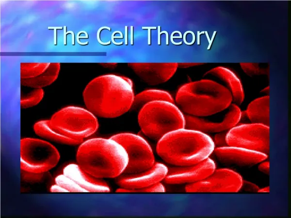
The Cell Theory Chapter 2
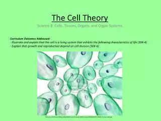
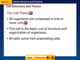
The Cell • All living things are made of cells. • Cells are the smallest living unit of life. • Each cell performs the necessary functions to sustain life. • Cells can replicate themselves. Cancer occurs when cells replicate haphazzardly. • Cellular function is carried out by organelles.
Cellular reactions are mediated (controlled) by ENZYMES. • Enzymes speed up chemical reactions. • All the chemical reactions in the body make up its METABOLISM. • There are 10 organelles that you are responsible for.
Plasma Membrane • Outer covering of the cell. • Also called the PLASMALEMMA. • Separates the intracellular (inside) fluid and the extracellular (outside) fluid. • Double layer of lipids (fat) with protein molecules between the layers. Fluid Mosaic Model
Polar Head (like water) • Non-polar tails (do not like water) • Proteins within the membrane. They give support to the membrane and prevent it from collapsing. • Carbohydrates are attached to the integral proteins. These form the GLYCOCALYX.
The glycocalyx is a sticky coat that allows the cell to bind to other cells. • The glycocalyx also serves as a marker. i.e.: certain antibiotics recognize the glycocalyx and attach to the cell.
Functions of the Plasma Membrane • Allows entry into the cell (Endocytosis) Phagocytosis – cell eating Pinocytosis – cell drinking Receptor-Mediated - hormones • Allows movement out of cells (Exocytosis)
Cytoplasm • Cytosol is a gel like substance which holds all the internal cellular organelles. • Contains ions, water, and enzymes.
THE ORGANELLES • Perform all the cellular functions. • “Little Organs”
MITOCHONDRIA • Power plant of the cell • Produces energy
RIBOSOME • 2 parts that make proteins. Ribosome RNA
ENDOPLASMIC RETICULUM • Subway system of the cell. • Network of membrane-walled tubes that twist through the cytoplasm. • ROUGH E.R. – have ribosomes attached. • SMOOTH E.R. – do not have ribosomes attached.
GOLGI APPARATUS • Stacks of disk shaped membranes. Sort Package proteins made in the ER Process
LYSOSOMES • Sacs containing digestive enzymes that can break down almost all types of biological materials. CELLULAR DRAIN-O
PEROXISOMES • Contain oxidases (use oxygen to neutralize aggressive compounds known as free radicals). • Free radicals can damage cellular proteins, membranes, and DNA if left to accumulate.
CYTOSKELETON • System of complex rods that run throughout the cytoplasm. • Microtubules • Microfilaments
CENTROSOME • Contains a matrix and an inner pair of centrioles which are important in cellular division.
NUCLEUS Membrane bound organelle which is the control center for all cellular activity.. Contains DNA – produce instructions for protein synthesis. Contain chromosomes Produce ribsomes in the nucleolus
Related Clinical Terms • NECROSIS – cellular death due to disease or injury. • HYPERTROPHY – increase in size. i.e. Muscle cells become hypertrophic in response to exercise.
Rough Endoplasmic reticulum Cell membrane Golgi apparatus Nuclear membrane nucleus nucleolus mitochondria Smooth Endoplasmic reticulum
TISSUES Chapter Four
OBJECTIVES for Tissues • Be able to list the four basic tissue types and give examples of each. • Describe the functions of each tissue type. • Describe the cellular components of each tissue type. • Describe and identify the morphology of epithelial tissue types.
Tissues • Cells do not operate independently. • Related cells work and operate together in organized groups. The bottom line: Tissues are clubs of cellular organization.
Four Types of Tissues • 1. Epithelial Tissue • 2. Connective Tissue • 3. Muscle Tissue • 4. Nervous Tissue
EPITHELIAL TISSUE Covers the body surface or Lines a body cavity
Epithelial Tissue • Occurs at the interface of two different environments. i.e. The epidermis is between the inside and the outside of the body. • Protects the body by detecting harmful stimuli. i.e. Receptors for pain are found within the skin.
Secretion release of molecules from the cell • Absorption bringing small molecules into the cell.
Ion transport – moves ions (charged molecules) across a membrane. • The epithelium filters fluids that cross the barrier.
Characteristics of Epithelial Tissue 1. Cellularity – composed almost entirely of cells. 2. Specialized Contacts – connects adjacent cells. • gap junctions, tight junctions, desmosomes 3. Polarity – under surface called the “basement membrane”. 4. Avascular – lacks blood vessels. Receives nutrients through the underlying connective tissue
Shapes of Epithelial Tissue • Simple Epithelium – one layer • Stratified Epithelium – more than one layer • Squamous – cells are wider than tall • Cuboidal – cells are just about as tall as wide • Columnar – cells are taller than wide.
Pseudostratified Ciliated Columnar Epithelium Pseudostratified ciliated columnar epithelium with pale goblet cells. The different levels of nuclei are clearer here. Again, notice the wavy-looking cilia
Stratified squamous epithelium with beginning surface cornification. This section is from thin skin, which has a dry surface covered with dead cells. Notice how flat the surface cells are and how dark and pyknotic (degenerative) their nuclei have become. Again, notice the distinct row of basal cells.
CONNECTIVE TISSUE • Types of connective tissue Fat Cartilage Ligaments and Tendons Bone Blood Collagen
Functions of Connective Tissue • 1. Support and bind other tissues. (tendons, ligaments) • 2. Hold body fluids (ground substance). • 3. Defend against infection - mast cells - macrophages - plasma cells - neutrophils • 4. Store nutrients as fat.
A stretched preparation of areolar connective tissue. The pink fibers of different thicknesses are collagenous (or white) fibers. The dark, thin, more tortuous fibers are elastic (or yellow) fibers. Most of the nuclei belong to fibroblasts.
Fat cells -- note nucleus and rim of cytoplasm pushed to one side by the accumulation of fat. The lipid itself has been dissolved out in fixation. In the center of the picture, in the space bounded by the four large fat cells, there is a small, round cross-cut of a capillary with a dark, shrunken red blood cell inside.
BLOOD - Neutrophil - Erythrocytes 1 – Erythrocyte 2 - Neutrophil
Muscle Tissue • Brings about body movement. • Moves by shortening. • Three Types: 1. skeletal – pulls on long bones. Striated. 2. cardiac – only in the heart. 3. smooth – no visible striations. Found in walls of hollow visceral organs such as digestive system, urinary organs, blood vessels, and uterus. Mostly involuntary.
Skeletal Muscle Fibers 1 - Multinucleated skeletal muscle fibers
Smooth Muscle Tissue 1 – Smooth muscle fibers 2 – Artery endothelium 3- Artery Lumen