MELANOMA
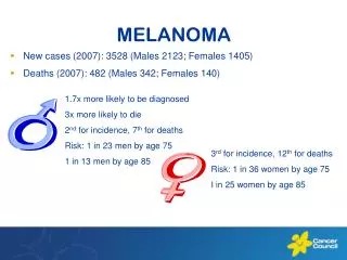
MELANOMA . Melanoma. Melanoma. Represents 4% of all cancers Most common malignancy in women aged 25-29. Incidence is rising faster than any other cancer Lifetime risk of developing melanoma now about 1 in 70 30% arise in existing nevi. Risk Factors. Fair skin Presence of atypical nevi

MELANOMA
E N D
Presentation Transcript
Melanoma • Represents 4% of all cancers • Most common malignancy in women aged 25-29. • Incidence is rising faster than any other cancer • Lifetime risk of developing melanoma now about 1 in 70 • 30% arise in existing nevi
Risk Factors • Fair skin • Presence of atypical nevi • Personal history of melanoma • Family history of atypical nevi or melanoma • History of blistering sunburn • Congenital nevi (incidence increases with increasing size)
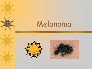
ABCDs of Melanoma Recognition • Asymmetry - one half of lesion does not look like the other • Borders - Scalloped border or focal extension into surrounding skin • Color variegation - varying hues and colors (black, brown, red, blue and white • Diameter - >6mm
Superficial Spreading Melanoma • Most common subtype accounting for 70-80% of melanomas • F > M mostly Caucasians • Most often on trunk and extremities • Tend to be > 6mm • Spread laterally for years before developing nodules
Nodular Melanoma • Account for 10-15% of melanomas • M = F • Most often found on the extremities • Evolve over months and extend vertically with little lateral spread
Lentigo Maligna and Lentigo Maligna Melanoma • Account for 5-10% of melanomas • Arise most often on the face, neck or dorsal arms in older Caucasians • M = F. Develop over years or decades • Lentigo Maligna represents in situ lesion and 5% progress to invasive LMM
Amelanotic Melanoma • Descriptive term for non-pigmented melanoma • Any type can be amelanotic • 2% of all melanomas are amelanotic • Malignant cells produce little or no pigment • Poorer prognosis due to delayed diagnosis
Acral Lentiginous Melanoma • Accounts for 7% of melanomas • Occurs on hands, feet, nails and mucous membranes • Most common melanoma in blacks and asians > 50% • Least common in Caucasians • Leading cause of skin cancer deaths
Acral Lentiginous Melanoma • Most patients are between 60 -70 • Usually slow growing • Often difficult to detect early which leads to poor prognosis • Often begins as a brown/black streak under a nail or bruise like area on acral skin
Acral Lentiginous Melanoma • Diagnosis is by clinical suspicion and tissue biopsy • Treatment is surgical excision with appropriate margins and in some cases requires amputation • Additional studies(PET, CT, SNL) are performed if indicated
Workup • All suspicious lesions should be biopsied. This does not increase the risk of metastases. • Excisional or punch biopsy allows for accurate measurement of Breslow’s depth. Never shave biopsy a suspected melanoma. • Breslow’s depth, ulceration and mitotic rate are most important features histologically • Sentinel lymph node biopsy should be done on all melanomas >=1mm • For patients with positive sentinel nodes, PET/CT scanning should be performed to rule out distant metastases
Treatment • Wide local excision with adequate margins is treatment of choice • Appropriate margin is determined by Breslow’s depth • Melanoma in situ requires a margin of 0.5 cm • Melanoma up to 2.0 mm requires a 1.0 cm margin with full thickness dermis and subcutaneous fat • Melanoma > 2.0 mm requires a 2.0 cm margin with full thickness dermis and subcutaneous fat
Sentinel Node Biopsy • Lymphoscintigraphy procedure where a sulfur colloid tagged with technetium-99m is injected at the site of the initial tumor and followed to the draining lymph node basin • 15 minutes prior to dissection blue dye is injected at the same site. • The nodal basin is then dissected and inspected for those nodes that take up the dye and are shown by Geiger counter to have taken up the radioactive material. These are the sentinel node(s)
Sentinel Nodes • Once identified the sentinel nodes are removed and examined histologically for evidence of tumor spread • If sentinel nodes are positive full elective lymph node dissection may be done as well as further staging workup • If sentinel node(s) is negative no further workup is required
Adjuvant Therapy • ELND has shown no real increase in survival • Adjuvant therapy with interferon was often recommended for those with advance stages of disease but did not significantly increase survival • Interferon has numerous side effects (flu like symptoms) making it difficult to complete treatment
Adjuvant Therapy • Zelboraf (vemurafenib) - For patients with metastatic melanoma with tumors that express a gene mutation called BRAF V600E. • BRAF helps regulate cell growth. The variant of BRAF targeted by Zelboraf is a gene mutation that allows melanoma cancer cells to spread • Almost 50 percent of all melanoma tumors have the BRAF genetic mutation • Not yet clear how long Zelboraf can increase melanoma survival.
Adjuvant Therapy • Yervoy (ipilimumab) - For the treatment of late-stage, metastatic melanoma • Patients taking Yervoy survived an average of 10 months after starting treatment • A monoclonal antibody that blocks a crucial switch on immune cells called CTLA-4. Cancers use this switch to turn off the body's anticancer immune responses. • Nearly 13% of patients taking Yervoy had severe or fatal autoimmune reactions.
Melanoma Survival • Stage IA: The 5-year survival rate is around 97%. The 10-year survival is around 95%. • Stage IB: The 5-year survival rate is around 92%. The 10-year survival is around 86%. • Stage IIA: The 5-year survival rate is around 81%. The 10-year survival is around 67%. • Stage IIB: The 5-year survival rate is around 70%. The 10-year survival is around 57%. • Stage IIC: The 5-year survival rate is around 53%. The 10-year survival is around 40%.
Melanoma Survival • Stage IIIA: The 5-year survival rate is around 78%. The 10-year survival is around 68%. • Stage IIIB: The 5-year survival rate is around 59%. The 10-year survival is around 43%. • Stage IIIC: The 5-year survival rate is around 40%. The 10-year survival is around 24%. • Stage IV:The 5-year survival rate for stage IV melanoma is about 15% to 20%. The 10-year survival is about 10% to 15%.
Prevention Wear protective clothing Always use sunscreen SPF 30 - 45. Must block both UVA and UVB Avoid midday sun 10 am - 3 pm Self skin exams Avoid sunburn
Follow Up • All melanoma patients should have semi-annual skin examinations including lymph node assessment for 2-3 years followed by annual exams for their lifetime • Appropriate scanning should continue as indicated • All first degree relatives should have a baseline skin exam and annual exams for life