Download
1 / 87
870 likes | 1.36k Views
The complications of diabetes mellitus are far less common and less severe in people who have well-controlled blood sugar levels.Wider health problems accelerate the deleterious effects of diabetes. These include smoking, elevated cholesterol levels, obesity,highblood pressure, and lack of regular exercise.

E N D
Slide 1:COMPLICATIONS OF DIABETES
By Dr Bashir Ahmed Dar
Associate Professor of Medicine
Chinkipora Sopore Kashmir
Email-- drbashir123@gmail.com
Slide 2:Dr Bashir and Dr yashodhora leading group of medical students to meet noble prize winner in medicine at KL Malaysia
Slide 3:Precious moments with noble prize winner Prof Barry. J Marshall for Helicobacter pylori cause for peptic ulcer
Slide 9:My Home in Kashmir
Slide 15:MY HOME S
Slide 16:IN MY YOUTH
Slide 17:SON YAWER BASHIR
Slide 22:Complications of Diabetes Short term Complications
Hypoglycemia
Diabetic Ketoacidosis
Non Ketotic hyperosmolar diabetic coma
Lactic acidosis
Slide 23:Long term Complications 1. Dermopathy
2. Retinopathy
3. Gastroentropathy
4. Neuropathy
5. Nephropathy
6. Myopathy
7. Osteopathy
Slide 24:Cutaneous complications of Diabetes Bacterial infections
Cellulitis
Folliculitis
Slide 25:Cutaneous complications of Diabetes Fungal Infections
Tinea pedis (athlete�s foot)
Tinea cruris (jock itch)
Slide 26:Cutaneous complications of Diabetes
Onychomycosis (fungal infection of the nails)
Slide 27:Cutaneous complications of Diabetes
Yellow Nails
Slide 28:Cutaneous complications of Diabetes Itching
Often a result of yeast infection, dry skin, and/or poor circulation
Slide 29:Cutaneous complications of Diabetes Digital Sclerosis
Tight, thick, waxy skin most commonly on the back of the hand
Finger joints can become stiff
Slide 30:Cutaneous complications of Diabetes Acanthosis Nigricans
Hyperpigmented plaques.
Most commonly found in armpits, neck, and groin
Slide 31:Acanthosis Nigricans
Slide 32:Acanthosis Nigricans
Slide 33:Acanthosis Nigricans
Slide 34:Cutaneous complications of Diabetes Diabetic Dermopathy.
Light brown, scaly, atrophic patches
Usually occur on the shins.round or oval, reddish-brown, scaly papules and plaques, ranging in size from 0.5 cm.Lesions do not itch, hurt, or open up.
Slide 35:Diabetic Dermopathy
Diabetic Dermopathy
Slide 36:Diabetic Dermopathy
Diabetic Dermopathy
Slide 37:Cutaneous complications of Diabetes Necrobiosis Lipoidica Diabeticorum
Similar to diabetic dermopathy but spots are fewer in number and larger and deeper
Can be itchy, painful, and difficult to treat
Slide 38:Cutaneous complications of Diabetes (Granuloma Annulare)
Slide 39:Cutaneous complications of Diabetes Pigmented Purpura
Brown to red macules and patches often coexists with diabetic dermopathy.
Slide 40:Cutaneous complications of Diabetes Diabetic blisters Bullosis Diabeticorum
Most commonly on the backs of fingers, hands, toes, feet, and sometimes on legs or forearms
Look like burn blisters
Slide 41:Bullosis Diabeticorum Bullae are blisters spontaneously appearing from normal skin. They are usually 0.5 to several centimeters in size, and contain a clear, sterile, viscous fluid
Slide 42:Bullosis Diabeticorum (cont.) These blisters sometimes are hemorrhagic and may heal with scarring and atrophy
Slide 43:Bullosis Diabeticorum (cont.)
Slide 44:Cutaneous complications of Diabetes Eruptive Xanthomatosis
Firm, yellow, pea-like enlargements in the skin
Most often on backs of hands, feet, arms, legs, and buttocks.
Slide 45:Cutaneous complications of Diabetes
Diabetic Foot Ulcers
Slide 46:Cutaneous complications of Diabetes Peripheral Vascular Disease
Diabetics are more prone to develop atherosclerosis, which can result in peripheral vascular disease Claudication
Delayed healing
Cold, pale, hairless legs and feet
Thick nails
Ulcers
Slide 47:Cutaneous complications of Diabetes
Gangrene
Slide 48:Necrobiosis Lipoidica Diabeticorum Presents with well-circumscribed erythematous papules, which develop into large, irregularly delineated plaques with a waxy, yellow center
Slide 49:Necrobiosis Lipoidica Diabeticorum The epidermis becomes thin and transparent, allowing underlying vasculature to become visible
The involved peripheral tissue is slightly raised and has a reddish-blue color
About 85% of NLD cases occur on the legs bilaterally
Lesions can also appear on the face, scalp, hands, forearms, or abdomen
Slide 50:Necrobiosis Lipoidica Diabeticorum (cont.)
Slide 51:Necrobiosis Lipoidica Diabeticorum (cont.)
Slide 52:Granuloma Annulare Granuloma annulare is identified by its characteristic annular or arciform plaques that begin as flesh-colored, red, or reddish-brown papules symmetrically spread across the upper trunk, neck, arms, and occasionally the legs
Slide 53:Granuloma Annulare (cont.)
Slide 54:Granuloma Annulare (cont.)
Slide 55:GIT COMPLICATIONS OF DIABETES The entire GI tract can be affected, including the mouth, esophagus, stomach, small intestine, colon, liver, and pancreas, leading to a variable symptom complex.
Gastroparesis leads to
Delayed gastric emptying
nausea, vomiting, bloating, postprandial fullness, or upper abdominal pain.
Intestinal enteropathy (which can cause diarrhea, constipation, and fecal incontinence)
Slide 56:GIT COMPLICATIONS OF DIABETES Esophageal manifestations of diabetic neuropathy, including abnormal peristalsis, spontaneous contractions
Impaired lower esophageal sphincter tone, result in heartburn and dysphagia
Slide 57:GIT COMPLICATIONS OF DIABETES Diabetes causes nonalcoholic fatty liver disease.
Nonalcoholic fatty liver disease is generally diagnosed because of persistent elevation in hepatic transaminase levels.
Diabetes is more common in patients with hepatitis C infection than in the general population.
Slide 58:GIT COMPLICATIONS OF DIABETES Causes of cirrhosis linked to diabetes include nonalcoholic fatty liver disease, hemochromatosis, and hepatitis C infection.
OHD can cause hepatotoxicity.
Slide 59:DIABETIC RETINOPATHY
Slide 60:DIRECT OPHTHALMOSCOPY Examination of the fundus of the eye
To screen for Diabetic Retinopathy
After dilatation of both eyes with 0.5% tropicamide
Slide 61:Normal fundus views of Right and left eye
Slide 62:View of the retina through an ophthalmoscope
Slide 63:View of the retina through an ophthalmoscope
Slide 64:View of the retina
Slide 65:Epidemiology of DR RISK of developing DR:
Type I or IDDM � 70%
Type II or NIDDM - 39%
Slide 66:Classification of DR Non-proliferative DR (NPDR)
Mild to moderate
Severe
2. Proliferative DR (PDR)
Slide 67:Classification of DR III. Diabetes also gives rise to
Clinically significant macular oedema (CSME)
which may exist by itself or along with NPDR and PDR
Slide 68:At least one microaneurysm � is the earliest clinically detectable lesion in MNPDR
and/or dot and blot hemorrhages in at least 1 quadrant
Hard or soft exudates (Cotton wool spots)
Venous beading
Slide 70:Mild and Moderate Non- proliferative DR was previously known as Background DR
Slide 71:Severe NPDR Any one of the following 3 features should be present
Microaneurysms and intraretinal hemorrhages in all 4 quadrants
Venous beading in 2 or more quadrants
Moderate IRMA in at least 1 quadrant
Known as the 4-2-1 rule
Slide 72:Severe NPDR Any one of the following 3 features should be present
Microaneurysms and intraretinal hemorrhages in all 4 quadrants
Venous beading in 2 or more quadrants
Moderate IRMA in at least 1 quadrant
Known as the 4-2-1 rule
Slide 74:Clinically significant Macular Oedema Retinal oedema close to fovea
Hard exudates close to fovea
Presents with dimness of vision
By itself or along with NPDR or PDR
Slide 75:CSME � Hard exudates close to fovea and associated retinal thickening
Slide 76:Proliferative DR (PDR) Characterized by Proliferation of new vessels from retinal veins
New vessels on the optic disc
New vessels elsewhere on the retina
Vascular Endothelial Growth Factor (VEGF) is produced by hypoxic retina
VEGF stimulates the growth of shunt and new vessels
Slide 78:Proliferative DR
Slide 79:COMPLICATIONS OF DIABETIC RETINOPATHY Vitreous hemorrhage
Tractional retinal detachment
Rubeosis Iridis
Glaucoma
Blindness
Slide 80:Vitreous hemorrhage is the extravasation of blood into one of the several potential spaces formed within and around the vitreous body. This condition may result directly from retinal tears or neovascularization of the retina, or it may be related to bleeding from preexisting blood vessels in these structures
Slide 81:The vitreous body occupies the entire space of the cavity of the eyeball comprised between the posterior surface of the lens and the retina. The clear colorless transparent jelly that fills the eyeball posterior to the lens
Slide 82:Retinal detachment
Slide 83:Retinal detachment Retinal detachment is a separation of the light-sensitive membrane in the back of the eye (the retina) from its supporting layers
Slide 84:Rubeosis Iridis Rubeosis iridis is a condition characterized by a new formation of vessels and connective tissue on the surface of the iris
Slide 85:Neovascular Glaucoma Complication of rubeosis iridis
New vessels cause angle closure
Mechanical obstruction to aqueous outflow
Intra ocular pressure rises
Pupil gets distorted as iris gets pulled
Eye becomes painful and red
Loss of vision
Slide 86:Blindness Non-clearing vitreous hemorrhage
Neovascular glaucoma
Tractional retinal detachment
Macular ischemia
Slide 87:Clinically significant macular oedema Give laser photocoagulation to minimise risk of visual loss and also given in case of PDN
It converts hypoxic retina (which produces ANGIOGENIC factors) into anoxic retina
Slide 88:Screening protocol for Diabetic retinopathy Screening once in a 1 year
Diabetics with normal fundus
Mild NPDR
Screening once in 6 months
Moderate NPDR
Slide 90:UROGENITAL COMPLICATIONS OF DIABETES Erectile dysfunction (ED).
Although most men do encounter trouble having an erection from time to time, the problem is not generally thought to be ED unless the symptoms are consistent for 3 months or more.
Slide 91:UROGENITAL COMPLICATIONS OF DIABETES Retrograde Ejaculation
Retrograde ejaculation is a condition in which part or all of a man�s semen goes into the bladder instead of out the tip of the penis during ejaculation. Retrograde ejaculation occurs when internal muscles, called sphincters, do not function normally. A sphincter automatically opens or closes a passage in the body.
Slide 92:UROGENITAL COMPLICATIONS OF DIABETES With retrograde ejaculation, semen enters the bladder, mixes with urine, and leaves the body during urination without harming the bladder. A man experiencing retrograde ejaculation may notice that little semen is discharged during ejaculation or may become aware of the condition if fertility problems arise. Analysis of a urine sample after ejaculation will reveal the presence of semen.
Slide 93:UROGENITAL COMPLICATIONS OF DIABETES Sexual problems in women may include
Decreased vaginal lubrication, resulting in vaginal dryness
Uncomfortable or painful sexual intercourse
Decreased or no desire for sexual activity
Decreased or absent sexual response in both sexes
constant or occasional inability to reach orgasm.
Slide 94:UROGENITAL COMPLICATIONS OF DIABETES Common bladder problems in men and women with diabetes include the following:
Overactive bladder. Damaged nerves may send signals to the bladder at the wrong time, causing its muscles to squeeze without warning. The symptoms of overactive bladder include
urinary frequency�urination eight or more times a day or two or more times a night
Slide 95:UROGENITAL COMPLICATIONS OF DIABETES urinary urgency�the sudden, strong need to urinate immediately
urge incontinence�leakage of urine that follows a sudden, strong urge to urinate.
Slide 96:UROGENITAL COMPLICATIONS OF DIABETES Poor control of sphincter muscles. Sphincter muscles surround the urethra�the tube that carries urine from the bladder to the outside of the body�and keep it closed to hold urine in the bladder. If the nerves to the sphincter muscles are damaged, the muscles may become loose and allow leakage or stay tight when a person is trying to release urine.
Slide 97:UROGENITAL COMPLICATIONS OF DIABETES Urine retention may also lead to overflow incontinence�leakage of urine when the bladder is full and does not empty properly.
Symptoms of urinary tract infections can include
a frequent urge to urinate
pain or burning in the bladder or urethra during urination
cloudy or reddish urine
in women, pressure above the pubic bone
in men, a feeling of fullness in the rectum
Slide 98:MUSCULOSKETAL COMPLICATIONS IN DIABETES
Slide 99:Diabetic Muscle Infarction
Rare
Painful muscle swelling, usually in thigh
Mass expands over days to weeks
Slide 100:Neuropathic Arthropathy(Charcot Joint) First described in 1868 by Jean Martin Charcot in patients with tabes dorsalis
Destructive arthropathy in diseases which impair sensory function, but maintain normal motor function
Slide 101:Charcot Joint Most common in MTPs, tarso-metatarsals, tarsus, ankle and interphalangeal joints
Single, painless, swollen, deformed joint in setting of peripheral neuropathy
Periarticular soft tissues loosen thereby causing joint laxity and subluxation
Repetitive microtrauma with weight bearing damages the joint
Slide 103:Hand Abnormalities Carpal Tunnel Syndrome
Dupuytren�s contracture
Flexor tenosynovitis
Limited joint mobility
Each condition present in ~ 20% patients with diabetes
Slide 105:Dupuytren�s Contracture Fibrosis in and around the palmar fascia with nodule formation
Contraction of the palmar fascia causes flexion contractures of digits
The 3rd and 4th finger most commonly effected in patients with diabetes, compared to the 5th finger in patients without diabetes
Present in 15-40% of patients with diabetes
Prevalence increases with age
Slide 107:Adhesive capsulitis ( frozen shoulder) Progressive painful restriction of shoulder movement
Joint capsule adheres to humeral head
3 phases: painful, adhesive, resolution
10-30% in diabetics, 2-10% in controls
17% patients with adhesive capsulitis have diabetes
Associated with age and duration of diabetes
Slide 108:Limited Joint mobility Diagnosis
�prayer sign�
�table top test�
To differentiate from Dupuytren�s:
Limited joint mobility usually involves 4 fingers
Absence of taut fibrotic bands
Slide 111:Diabetic Sclerodactyly Thickening and waxiness of skin
Usually on dorsa of fingers
Associated with limited joint mobility
Similar to skin changes of scleroderma
(absent antibodies, Raynaud�s, calcinosis, ulceration, tapering)
Slide 112:Diabetic neuropathy There are two types of diabetic neuropathy
Diffuse peripheral neuropathy primarily affects the limbs, damaging the nerves of the feet and hands.
Focal�or localized neuropathy affects specific nerves, most commonly in the torso, leg, or head.
Slide 113:Autonomic neuropathy Autonomic neuropathy is the other form of diffuse neuropathy and it affects the heart and other internal organs.
Slide 114:Autonomic neuropathy Diabetic neuropathy can lead to
muscular weakness,
Loss of feeling or sensation,
and loss of autonomic functions such as
Digestion,
Erection,
Bladder control, and sweating
Fecal incontinence)
Slide 115:Autonomic neuropathy Stomach disorders, due to the impaired ability of the stomach to empty (gastric stasis)
Nausea,
Vomiting
and bloating
Diarrhea
constipation
Slide 116:Autonomic neuropathy Lightheadedness
and fainting spells
Loss of appetite
Abdominal swelling
Heat intolerance
Impotence
Slide 117:Autonomic neuropathy Dizziness with standing
Exercise intolerance
Orthostatic hypotension
Any cranial nerve can be affected as focal neuropathy
Slide 118:Diabetic neuropathy In severe diabetic neuropathy loss of sensation can lead to injuries that are unnoticed, progressing to infections, ulceration and possibly amputation.
Slide 119:Diabetic peripheral neuropathy Sensory ? Motor (myelin)
Characteristic features of Peripheral Neuropathy are
Bilateral, symmetrical
Progressive
Paraesthesias, pain, muscle atrophy
Glove and stocking type usually affects distal parts of limbs then ascend upwards
Slide 121:Chronic Polyneuropathy Claw foot � Dermopathy & Neuropathy
Slide 122:Diabetic Amyotrophy Painful muscle wasting
Slide 123:Diabetic Neuropathic ulcer
Slide 124:Neuropathic ulcer Etiology:
peripheral sensory neuropathy, Trauma & deformity.
Factors:
Ischemia, callus formation, and edema.
Slide 125:Neuropathic ulcers FEATURES:
Painless, surrounded by callus
At pressure points.
associated with good foot pulses
May not be associated with gangrene
Slide 126:Nephropathy An angiopathy of glomeruli
Is a micro vascular complication of diabetes marked by albuminuria and a deteriorating course from normal renal function to end stage renal failure. ESRD
Slide 127:Risk factors for nephropathy Hypertension
Hyperglycemia
Microalbuminuria
Ethnicity
Male gender
Family history
Cigarette smoking
Slide 128:Nephropathy The syndrome was discovered by British physician Clifford Wilson (1906�1997) and German-born American physician Paul Kimmelstiel (1900�1970) and was published for the first time in 1936.
Slide 129:Nephropathy Usually manifests 15�25 years after diagnosis of diabetes .
The disease is progressive and may cause death two or three years after the initial lesions, and is more frequent in men.
Slide 130:Nephropathy The glomeruli and kidneys are typically normal or increased in size initially, thus distinguishing diabetic nephropathy from most other forms of chronic renal insufficiency, wherein renal size is reduced (except renal amyloidosis and polycystic kidney disease).
Slide 131:Approximately 25% to 40% of patients with DM 1 ultimately develop diabetic nephropathy (DN), which progresses through five predictable stages.
Slide 132:Increased demand upon the kidneys is indicated by an above-normal glomerular filtration rate (GFR).
Hyperglycemia leads to increased kidney filtration
This is due to osmotic load and to toxic effects of high sugar levels on kidney cells
Increased Glomerular Filtration Rate (GFR) with enlarged kidneys
All this results in nephromegaly
Slide 133:Clinically silent phase with continued hyper filtration and hypertrophy .The GFR remains elevated or has returned to normal, but glomerular damage has progressed and leads to next stage that is chracterised by microalbuminuria with excretion of albumin in the range of 30-300 mg/day .
Normal persons excrete less than 30mg/day. at this stage the disease process is probably reversible.
Slide 134:
Significant microalbuminuria will progress to end-stage renal disease (ESRD).
Therefore, all diabetes patients should be screened for microalbuminuria on a routine basis. 134
Slide 135:In this stage albumin is more than 300 mg in a 24 hour period. The urine becomes "dipstick positive,�
Albumin more than 300 mg/24 hour is called macroalbuminuria (defined as >300 mg/day (200 microgram/min). from this stage the disease is irreversible and a steady decline in glomerular filtration occurs at a rate of 1 ml/minute per month. the stage of macroalbuminuria may progress to nephrotic syndrome.
If proteins are more than 3gm/24 hours then it results in nephrotic syndrome.
Slide 136:Hypertension (high blood pressure) typically develops during stage 3.
Basement membrane thickening occurs due to deposition of AGEP
Slide 137:Glomerular damage continues, with increasing amounts of protein albumin in the urine.
The kidneys� filtering ability has begun to decline steadily, and blood urea nitrogen (BUN) and creatinine (Cr) has begun to increase.
Slide 138:With further progression the azotaemia develops and progression to renal failure and uraemia is inevitable.
The glomerular filtration rate (GFR) decreases about 10% annually. Almost all patients have hypertension at stage 4.
Slide 139:GFR has fallen to <10 ml/min and renal replacement therapy (i.e., haemodialysis, peritoneal dialysis, kidney transplantation) is needed.
Slide 140:Glomerular Histology:�
The glomerular capillary wall is composed of an endothelial cell layer (blood side), a thick basement membrane, and epithelial cell layer (urine side).
�
(i) Glomerular Endothelium
The glomerular endothelium is fenestrated. The fenestrae (0.07 to 0.1 mm-micrometers- in maximal diameter) allow the passage of electrolytes, proteins, and globulin.
However, platelets (3 mm), red cells (7 mm) and neutrophils (15 mm) can't pass through the endothelial layer.
Slide 142:(ii) Glomerular Basement Membrane (GBM):
The GBM is a tri-laminar structure, 0.3 microns in thickness, composed of collagen, proteoglycans and laminin.
It is product of the fusion of the endothelial and epithelial basement laminae.
The dense central GBM area, or lamina densa, is due to the overlapping of the two laminae. �
Around 50% of the GBM is collagen IV.
Slide 144:The negative charge of the GBM has been attributed to the presence of the heparan sulphate proteoglycan (HSPG) called perlecan.
These negatively charged molecules are geometrically arranged in clusters separated by about 0.003 �m from each other.
This anionic molecular sieve restricts the passage of molecules according to size and charge.
Water, salts, glucose, amino acids and neutral, or cationic, molecules with radii less that 0.0035 �m are filtered with relative ease.
The albumin molecule measures 0.0035 �m and is negatively charged. Therefore its filtration is restricted.
Slide 145:Diabetic nephropathy The negative charge of the GBM has been attributed to the presence of the heparan sulphate proteoglycan (HSPG) called perlecan.
These negatively charged molecules are geometrically arranged in clusters separated by about 0.003 �m.
The albumin molecule measures 0.0035 �m and is negatively charged. Therefore its filtration is restricted.
Slide 146:Diabetic nephropathy Presence of protein in the urine is a sign that either the charge or the distance between the anionic clusters, or both, are pathologically altered.
The presence of red cells in the glomerular urine, is certain indication of GBM ruptures.
Slide 147:Mesangium Mesangial Matrix is a cellular network like a thin membrane along inner side of basement membrane composed of different types of collagens (I, III, IV), laminin , entactin and proteoglycans.
it is surrounding the glomerular capillaries and keeps the glomerular capillaries together.
Slide 148:Mesangium Mesangium is composed of mesangial cells type I and II, and other tissue matrix.
Mesangial type I cells are monocytes with phagocytic functions. These cells can extend cytoplasmic projections into the glomerular capillary.
Slide 150:Mesangium They also "clean" the mesangium of materials that leak from the capillary lumen into the matrix. These cells are stimulated by cytokines to produce free radicals and cytotoxic peptides.
Mesangial type II cells are myofibroblasts with the ability to contract upon ADH and angiotensin stimulation.
Their contraction causes a reduction of the effective glomerular filtration area.
Slide 152:Diabetic nephropathy Three major histological changes occur in the glomeruli of persons with diabetic nephropathy.
1.Mesangial expansion is directly induced by hyperglycemia
perhaps via increased matrix production or glycosylation of matrix proteins. the cells mesangical that surrounds to glomerular vessels increases as a result of material similar to material of basement membrane.
2.GBM thickening
3.Glomerular sclerosis is caused by intraglomerular hypertension (induced by renal vasodilatation or from ischemic injury induced by hyaline narrowing of the vessels supplying the glomeruli).
Slide 153:Diabetic nephropathy Glomerular Hyper filtration
Glucose provides an osmotic diuretic effect
Result is increased renal filtration. Kidney responds with hypertrophy of epithelium and endothelium leading to glomerular hypertrophy.
Glomerular pressure increases
Result is premature glomerulosclerosis
Slide 154:Diabetic nephropathy Humoral Imbalances in DM Nephropathy
Insulin Deficiency
Elevated Glucagon Concentrations
Increased Transforming Growth Factor (TGF)-�
Increased angiotensin II
Abnormally regulated thromboxanes and endothelins
Abnormal insulin like growth factor (IGF)-1
Elevated platelet derived growth factor (PGDF)
Slide 155:Diabetic nephropathy Role of TGF-�
Stimulates extracellular matrix synthesis,tubuloepithelial hypertrophy, basement membrane thickening, proliferation of fibroblasts
Angiotensin II and Thrombospondin (TSP1) can both stimulate the production of transforming growth factor-�.
Slide 157:Diabetic nephropathy Ultra structural changes of the glomerular basement membrane in diabetic nephropathy reveals
The normal human GBM shows pores.
The diameter of these pores was slightly smaller than that of human albumin molecules.
The GBM in patients with diabetic nephropathy shows formation of unknown cavities and tunnel structures in the thickened GBM
In some portions, these cavities presented a honeycomb-like appearance.
Slide 158:Diabetic nephropathy The diameters of the cavities and tunnels are far larger than the dimensions of albumin molecules.
These enlarged structures are believed to allow serum protein molecules to pass through the GBM from the capillary lumen to the urinary space.
These results suggest that the cause of massive proteinuria in diabetic nephropathy is the disruption of the size barrier of the GBM.
Slide 159:Diabetic nephropathy Non-enzymatic glycosylation of proteins and lipids exposed to sugars form reversible Schiff bases or Amadori products.
Later, through further molecular rearrangements, irreversible advanced glycosylation end products (AGEs) are formed that appear as nodular or diffuse deposits.
and contribute to basement membrane (and mesangial) thickening-glomerulosclerosis, change permeability (leading to proteinuria, retinal hard exudates and microhaemorrhages).
Slide 160:Diabetic nephropathy Thickening of basement membranes is thus partly due to glycosylation of membrane proteins or entrapment of glycosylated serum proteins into basement membrane.It is evident that AGEs can interact with cell functions, tissue remodelling and inflammatory reactions in several different ways.
When Ang II is increased, constriction of efferent than afferent arterioles occurs that increases glomerular filtration rate and raises intraglomerular pressure, causing glomerular hypertension.
Sustained or severe increases in intraglomerular pressure can lead to GBM damage, glomerular endothelial dysfunction, and ultimately, extravasation of protein into Bowman�s capsule.
Slide 161:Diabetic nephropathy Attempts by the proximal tubules to reabsorb this filtered protein causes injury to the tubular cells, activates an inflammatory response, that create further stress on the already compromised glomerulus.
The resultant tubular inflammatory response and renal microvascular injury activate pathways that lead to fibrosis and scarring of both glomerular and tubular elements of the nephron ultimately leading to contracted kidneys.
Slide 163:Summary Pathological lesions in DM The Armani-Ebstein change (or Armani-Ebstein cells) consists of deposits of glycogen in the tubular epithelial cells (pars straight of proximal convoluted tubule and loop of Henle).
Slide 164:Summary Pathological lesions in DM Because most diabetics are treated before this stage, it is very rare to see it at the present time. It appears in decompensated diabetics with glycemia higher than 500 mg/dL and in the presence of severe glycosuria; it is a reversible alteration without functional manifestations.
Slide 165:Summary Pathological lesions in DM the basement of glomerular capillaries thicken and can obliterate the blood vessels.
The glomerular sclerosis is diffuse, but in 50% of cases it is accompanied with nodular sclerosis.
The nodular component denominated Kimmelstiel-Wilson nodules is pathognomic of diabetes.
Slide 167:Summary Pathological lesions in DM In short
Microangiopathy
Atherosclerosis
Diffuse glomerulosclerosis
or nodular diabetic glomerulosclerosis (Kimmelstiel Wilson nodules)
Tubulointerstitial fibrosis
Slide 169:Nodular Glomerulosclerosis � KW lesion.
Slide 170:Summary Pathological lesions in DM Arteriosclerosis and hyalinosis of afferent and efferent arterioles .
Necrotizing renal papillitis.
Infections &Pyelonephritis.
Nephrotic syndrome
End stage kidney
Slide 172:Summary Pathological lesions in DM A kidney biopsy confirms the diagnosis. However, your doctor can diagnose the condition without a biopsy if you meet the following three conditions:
1.Persistent protein in the urine
2.Diabetic retinopathy
3.No other kidney or renal tract disease
A biopsy may be done, however, if there is any doubt in the diagnosis
Slide 173:Treatment of nephropathy Factors that favor the regression of microalbuminuria include better blood sugar control, lower blood pressure, lower serum cholesterol and triglycerides, recent onset and lower levels of microalbuminuria, and less glomerular hyperfiltration.
Slide 174:Treatment of nephropathy Early screening
Spot urine albumin : creatinine ratio
24 hour urine collection
dipstick
Tight glycemic control
Slide 175:Treatment of nephropathy ACE inhibitors if creatinine less than 3mg/dl
Use ACEI as first line, if not tolerated, use ARB. Use the maximum dose as tolerated
If still hypertensive or proteinuric, consider using combination ACEI and ARB, or ACEI and diuretics
Slide 176:Treatment of nephropathy ACE inhibitors or ARB have a strong antiproteinuric effect apart from their antihypertensive actions
Increasing the dose of the ACEI or ARB beyond the optimum antihypertensive doses further reduces proteinuria
Antiproteinuric effect is enhanced by a low Na diet or diuretic
Slide 177:Treatment of nephropathy 0.8g/kg/day proteins
Antilipids
Thiazide diruritics
Vit E
Slide 178:Treatment of nephropathy Keep BP slightly less than 130/85 mmHg
Patients with CKD and > 1g proteinuria, BP goal should be < 125-130/75-80 mmHg
Slide 179:Complications of nephropathy Possible complications of diabetic nephropathy include:
hypoglycemia (from decreased excretion of insulin)(insulin isn't secreted by the kidneys)
rapidly progressing chronic kidney failure
end-stage kidney disease
Hyperkalemia
Nephrotic syndrome
Slide 180:Complications of nephropathy severe hypertension
complications of hemodialysis
complications of kidney transplant
coexistence of other diabetes complications
peritonitis (if peritoneal dialysis used)
increased ifections
Slide 181:Diabetic Gangrene
Slide 182:Blood vessel calcification:
Slide 183:Cataract
Slide 184:Acanthosis Nigricans
Slide 185:Acanthosis Nigricans
Slide 186:Label the diagram. 1.
2.
3.
4.
Slide 187:Infections in Diabetes: Decreased metabolism � low immunity.
Decreased function of lymphocytes & neutrophils � glycosylation.
Glycosylation of immune mediators. Ab
Capillary thickening � impaired inflammation.
Ischemia & infarctions.
Increased glucose (alone is not the cause*)
Diabetes ? State of immunosuppression.
Slide 188:Laboratory Diagnosis: Urine glucose - dip-stick �Screening
Random or fasting blood glucose (<11)
Fasting > 7mmol, Random >11mmol
If Fasting level is between 7-11 then OGTT
HbA1c - for follow-up, not for diagnosis
Fructosamine - for long term maintenance.
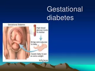
Slide 189:Gestational diabetes Gestational diabetes and impaired glucose tolerance (IGT) in pregnancy affects between of all pregnancies and both have been associated with pregnancy complications.
Slide 190:Fasting and 2 hours postprandial venous plasma sugar during pregnancy.
Slide 191:50-g oral glucose challenge The screening test for GDM, a 50-g oral glucose challenge, may be performed in the fasting or fed state. Sensitivity is improved if the test is performed in the fasting state .
A plasma value above one hour after is commonly used as a threshold for performing a 3-hour OGTT.
If initial screening is negative, repeat testing is performed at 24 to 28 weeks.
Slide 192:3 hour Oral glucose tolerance test Prerequisites:
- Normal diet for 3 days before the test.
- No diuretics 10 days before.
- At least 10 hours fast.
- Test is done in the morning at rest.
Slide 193:3 hour Oral glucose tolerance test Giving 75 gm (100 gm by other authors) glucose in 250 ml water orally
Slide 194:3 hour Oral glucose tolerance test Criteria for glucose tolerance test:
The maximum blood glucose values during pregnancy:
- fasting 90 mg/ dl,
- one hour 165 mg/dl,
- 2 hours 145 mg/dl,
- 3 hours 125 mg/dl.
If any 2 or more of these values are elevated, the patient is considered to have an impaired glucose tolerance test.
Slide 195:Affects of diabetes on pregnancy birth defects or be stillborn.
Infants of mothers with preexisting diabetes experience double the risk of serious injury at birth, triple the likelihood of cesarean delivery, and quadruple the incidence of newborn intensive care unit (NICU) admission.
Slide 196:Affects of diabetes on pregnancy Infants ,large for gestational age (macrosomic) and small for gestational age. Macrosomia in turn increases the risk of instrumental deliveries (e.g. forceps, ventouse and caesarean section) or problems during vaginal delivery (such as shoulder dystocia).
Slide 197:Affects of diabetes on pregnancy Neonates are also at an increased risk of low blood glucose (hypoglycemia), jaundice, high red blood cell mass (polycythemia) and low blood calcium (hypocalcemia) and magnesium (hypomagnesemia).GDM also interferes with maturation, causing dysmature babies prone to respiratory distress syndrome due to incomplete lung maturation and impaired surfactant synthesis
Slide 198:Affects of diabetes on pregnancy Studies have shown that the offspring of women with GDM are at a higher risk for congenital malformations.
women with GDM have a higher risk of preeclampsia
spontaneous abortion and congenital malformations in infants
Slide 199:Indications for detection of diabetes in pregnant women
Family history of Diabetes
Glucose in urine sample
History of unexplained prenatal loss
History of large baby
Slide 200:Indications for detection of diabetes in pregnant women History of congenitally malformation
Maternal obesity
Maternal age more than 25 years
Members of ethnic/racial group with high prevalence of Diabetes Mellitus
Slide 201:Management during pregnancy Quit smoking/alcohol
Home blood glucose monitoring
Diet control/Folate supplementation
Regular exercise
Fetal monitoring by ultrasound scan
Accurate insulin regimen
Slide 202:Diabetes and Dental health
Slide 203:The Importance of Dental Care With Diabetes Diabetes is a disease that can affect the whole body, including your mouth.
Dental care is particularly important for people with diabetes because they face a higher than normal risk of oral health problems due to poorly controlled blood sugars. The less well controlled the blood sugar, the more likely oral health problems will arise. This is because uncontrolled diabetes impairs white blood cells, which are the body's main defense against bacterial infections that can occur in the mouth.
Slide 204:What Dental Problems are You at a higher risk for? Dry mouth, xerostomia and salivary gland dysfunction
Caries
Increased susceptibility to bacterial, viral and fungal (that is, oral candidiasis) infections
Periapical abscesses
Periodontitis and loss of teeth
Lichen planus
Burning Mouth Syndrome
Gingivitis
Slide 205:Dry Mouth Uncontrolled diabetes can decrease saliva flow, resulting in dry mouth. Dry mouth can further lead to sores, ulcers, infections, and tooth decay.
Slide 206:Dry Mouth Leads to Ulcers and Sores
Slide 207:Cause difficulties in tasting, chewing, swallowing, and speaking
Increase your chance of developing dental decay and other infections in the mouth
Be a sign of certain diseases and conditions
Be caused by certain medications or medical treatments
Slide 208:Symptoms Include� A sticky, dry feeling in the mouth
Trouble chewing, swallowing, tasting, or speaking
A burning feeling in the mouth
A dry feeling in the throat
Cracked lips A dry, tough tongue
Mouth sores
An infection in the mouth
Decay, when there is not an adequate supply of saliva, the rate of tooth decay increases rapidly�
Slide 209:Symptoms Include� The average person creates around 1 Liter of saliva a day.�
If saliva production is reduced, an individual's oral bacteria levels can increase 10 times over normal levels.
Slide 210:Salivary gland dysfunction and Xerostomia There are reports of dry mouth complaints, which is known as xerostomia, as well as salivary hypofunction in patients with diabetes, which may be due to polyuria, or an underlying metabolic or endocrine problem.
When the normal environment of the oral cavity is altered because of a decrease in salivary flow or alteration in salivary composition, a healthy mouth can become susceptible to dental caries and tooth deterioration.
Slide 211:Treatment of Dry Mouth: a high-fluoride toothpaste like Colgate�s Prevident 5000+ to help reduce decay
Biotene, an oral rinse found over the counter which relieves of dry mouth.
Salagen ( Pilocarpine). Salagen pills have been shown to provide significantly increased saliva flow and relief of dry mouth. Since Salagen may cause fluctuations in blood pressure or heart rate, you should be closely supervised by an M.D.
Slide 212:Treatment of Dry Mouth: Chew gum or sour candy.� Look for sugarless gum or candy with Xylitol.
If you chew this gum for five minutes after every meal, studies show that you can reduce the incidence of tooth decay up to 62%. �
Slide 213:Dental Caries & Cavities It occurs when your teeth are frequently exposed to foods containing carbohydrates such as starches and sugars like soda pop, candy, cake and even sticky fruits.
Cavities have been identified
as a bacterial infection.
Bacteria inhabit the plaque and form up to 500 different
Slide 214:How Cavities Form Plaque interacts with food deposits on your teeth to produce acid that will slowly dissolve the calcium in your teeth. The surface of the tooth..."enamel" is 97% calcium, causing tooth decay and some of the other products cause gum disease and bad breath.
When enough calcium dissolves from the tooth surfaces, the surface breaks and forms a hole. That is how cavities form.� An active lesion demineralizes the tooth and can be diagnosed based upon color, surface texture and x-rays.�
White spots can be active lesions if they are not glossy, and feel rough to the explorer.
Slide 215:Cavities An area of decay may take as long as 6-8 years� or as short as 6 months to dissolve the outer layer (enamel) of the tooth.� If you have a "cavity" this outer layer has collapsed producing a hole that cannot repair itself.
Slide 217:Tooth decay & cavities Root cavities:
As you age, your gums can recede, leaving parts of your tooth root exposed. Since there is no enamel covering your tooth roots, these exposed areas easily decay. Most people over 60 have root cavities as a result of gum disease.
Slide 218:Tooth decay & cavities Gum recession has been found to occur more frequently and more extensively in moderate-and poorly-controlled diabetic patients because plaque responds differently, creating more harmful proteins in the gums.�
Slide 219:Tooth decay & cavities Repeated decay around existing fillings:
Decay can form around existing fillings and crowns. This is because these areas are not as smooth as a natural tooth surface and can decay easier.
Slide 220:Bacterial, Viral and Fungal Infections Thrush, or oral candidiasis, is an infection caused by a fungus that grows in the mouth. People with diabetes are at risk for thrush because the fungus thrives on high glucose levels in saliva.
Smoking, taking antibiotics often, or wearing dentures, (especially when they are worn constantly), can also lead to this fungal infection. Medication is available to treat this infection.
Good diabetic control, not smoking, and removing and cleaning dentures daily can help prevent thrush.
Slide 221:Thrush
Slide 222:Periapical Abscesses A dental abscess is an infection of the mouth, face, jaw, or throat that begins as a tooth infection or cavity.
Although these infections can be caused by poor dental health and can result from lack of proper and timely dental care, they may also occur in people with underlying autoimmune disorders and people who have other conditions that weaken the immune system (diabetes, post-radiation/chemotherapy cancer care & diabetes).
Dental abscesses can also be triggered by minor trauma in the oral cavity.
Slide 223:Periapical abcess
Slide 224:Periapical Abscess
Slide 225:Periodontitis Diabetics are more prone to the development of gum disease, (periodontal disease), from gingivitis that is caused by the presence of bacteria in plaque
Slide 226:Periodontitis Plaque is the white sticky film that accumulates on teeth both above and below the gum line that can harden into a rough yellow or brown deposit called tartar or calculus.
Slide 227:Any periodontal disease you may develop can be more severe and harder to control� Without regular dental checkups, periodontal disease may result if gingivitis is left untreated.� It can also cause inflammation and destruction of tissues surrounding and supporting teeth, gums, bone and fibers which hold the gums to the teeth.�Gum infections can make it hard to control blood sugar.�
Once a gum infection starts, it can take a long time to heal. If the infection is severe, teeth can loosen or even fall out.
Slide 229:Periodontitis Facts It has been shown that patients with type 2 diabetes are three times more likely to develop periodontal disease than are people without diabetes.
When people with diabetes smoke, they are 20 times more likely to develop periodontitis with loss of supporting bone than are those without diabetes.
One-third of people with DM have severe periodontal disease
Slide 230:Diabetes and Periodontitis
Periodontal disease has been proposed as the sixth complication of DM; the other five complications are retinopathy, neuropathy, nephropathy, cardiovascular disease and peripheral vascular disease
Slide 231:Lichen Planus Lichen planus is a relatively common, chronic mucocutaneous disease of unknown cause. It generally is considered to be an immunologically mediated process that involves a hypersensitivity reaction on the microscopic level.
In the mouth, it looks like lacy white patches on the inside of the cheeks or on the tongue.
Slide 232:Oral Lichen Planus
Slide 233:Causes of Lichen Planus
Slide 234:Causes of Lichen Planus The link with diabetes and oral lichen planus is more than likely an adverse affect of the drug therapy used to treat diabetes mellitus.
Slide 235:Burning Mouth Syndrome Patients with burning mouth or burning tongue syndrome usually exhibit no clinically detectable lesions, although the symptoms of pain and burning can be intense. The etiology of burning mouth is varied and often difficult to decipher clinically.
Burning mouth syndrome (BMS) is a complex, vexing condition in which a burning pain occurs on your tongue or lips, or over widespread areas involving your whole mouth, without any obvious reason.
Slide 236:BMS
Slide 237:BMS The main symptom of burning mouth syndrome is a burning sensation in your tongue, lips, gums, palate or throat. People with the syndrome may describe the sensation in the affected areas as hot or scalded, as if they had been burned with a hot liquid.
Other symptoms may include:
Dry mouth
Sore mouth
A tingling or numb sensation in the mouth or on the tip of the tongue
A bitter or metallic taste
Slide 238:Causes of BMS Dry mouth
Oral thrush
Geographic tongue
Depression
Vitamin deficiency anemia
Heartburn/GERD
Bruxism
Diabetes
Hypothyroidism
Menopause
Certain medications
Allergies
Slide 239:Gingivitis and Diabetes Gingivitis, a reversible condition, is characterized by inflamed and bleeding gums. Since it can be a precursor to chronic periodontitis, gingivitis requires treatment.
In gingivitis, periodontal disease is confined to the gingiva with no loss of junctional epithelial attachment. Gingivitis results from bacterial plaque accumulation at the gum margin and in the sulcus between the margin and the tooth.
Slide 240:Gingivitis and Diabetes Thickening of blood vessels is a complication of diabetes that may increase risk for gum disease.� Diabetes causes blood vessels to thicken, which slow the flow of nutrients to the mouth and slows the removal of harmful wastes away from the mouth.
Slide 241:Gingivitis and Diabetes When diabetes is poorly controlled, high glucose levels in mouth fluids may help germs grow and set the stage for gum disease.
Smoking increases the risk for gum disease.� If you are a smoker with diabetes, age 45 or older, you are 20 times more likely than a person without these risk factors to get severe gum disease, bone loss and tooth loss.
Slide 242:Gingivitis
Slide 243:You can do these simple things to help reverse gingivitis and prevent periodontal disease: Diet and exercise may be the most important changes that you can make to improve your quality of life and oral health.��
Brush your teeth after each meal.
Floss daily.
Get regular dental cleanings and check-ups.
Scrape your tongue with a tongue scraper.
Be sure both their medical and dental care providers are aware of your medical history and periodontal status.��
Be aware of your blood sugar levels, triglycerides and cholesterol levels and have them checked on a regular basis.� If your gums bleed while you are brushing your teeth or eating, or a bad taste stays in your mouth, go to the dentist.� Tell the dentist about any other changes you see, such as white patches, in your mouth.
Slide 244:When is the best time to receive dental care if you are a diabetic?
Slide 245:Best time to receive dental care Dental procedures should be as short and as stress free as possible.Make morning
appointments because blood glucose levels tend to be under better control at this time of day.
If you have a scheduled appointment, eat and take your medications as directed.
Test your blood sugar level and take your blood pressure and bring these results with you to our office.
Be prepared to update your health/dental history at each visit so we can provide you with the best possible care for your condition.
Slide 246:Best time to receive dental care Postpone non-emergency dental procedures if your blood sugar is not in good control.� However, abscesses should be treated right away.
See your dentist on a regular basis, every 3 to 4 months, for exams and cleanings. Keep the dentist informed of your health status and if you have any problems controlling your blood sugar.
Know that healing time will take longer due to your diabetic condition.
Slide 247:THANK YOU SO MUCH Trust the physician and the teacher, and drink his remedy in silence and tranquility. For his hand though heavy and hard is guided by tender hand of unseen. And the cup he brings, though it burn your lips has been fashioned of the clay which the potter have moistened with his tears and sacred feelings.