Download
1 / 56
560 likes | 692 Views
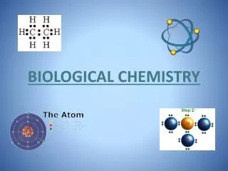
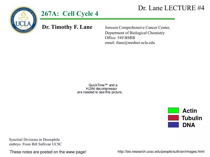
Dr. Lane LECTURE #4. 1. 267A: Cell Cycle 4. Dr. Timothy F. Lane Jonsson Comprehensive Cancer Center, Department of Biological Chemistry Office: 549 BSRB email: tlane@mednet.ucla.edu. Actin. Tubulin. DNA. Syncitial Divisions in Drosophila embryo. From Bill Sullivan UCSC.

E N D
Dr. Lane LECTURE #4 1 267A:Cell Cycle 4 Dr. Timothy F. Lane Jonsson Comprehensive Cancer Center, Department of Biological Chemistry Office: 549 BSRB email: tlane@mednet.ucla.edu Actin Tubulin DNA Syncitial Divisions in Drosophila embryo. From Bill Sullivan UCSC These notes are posted on the www page! http://bio.research.ucsc.edu/people/sullivan/images.html
Last time: We talked more about the regulation of the cell cycle by cyclins and cdks: i. That G1 passage required E2F transcription factors, but that these proteins were held in an inactive state by RB. ii. That cdk4/cycD phosphorylation of pRB is required to release E2F and activate S phase genes (including cycE). ii. That S phase entry required cycE/cdc2 kinase activity. iii. Derepression of Histone genes required cycE/cdc2 phosphorylation of pNPAT We discussed multiple levels of regulation for cyclins and cdks, including: i. Transcriptional control of synthesis (pRB represses cycE) ii. Phosphorylation (wee1 etc) / dephosphorylation (cdc25) iii. binding to CKIs (Far1, Sic, p15) iv. turnover (TODAY!) We discussed results from KO mice that indicate many cyclins and cdks are dispensable for simple proliferation in mammalian cells, but are required for regulation and inductive activities in complex organisms.
Goals: The context will be Mitosis. How M-phase is regulated/ended! Examine how cyclins are turned over in the context of M
Mitosis: G2 S G1 Image by Abby Marsh Image by Abby Marsh
Mitosis: G2 S G1 Image by Abby Marsh Image by Abby Marsh
Cohesins cyclin B deg. cdc2/cyclin B G2 Prophase Metaphase Anaphase Telophase G1 Mitosis: Active cyclinB/p34cdc2 is required to initiate mitosis.
Cohesins cyclin B deg. cdc2/cyclin B G2 Prophase Metaphase Anaphase Telophase G1 Mitosis: Active cyclinB/p34cdc2 is required to initiate mitosis. Exit from the mitotic state requires inactivation of the p34cdc2 kinase. and cyclin B must be degraded. Questions: What is the signal for cyclin B degradation? How is cyclin B degraded?
Cyclin B degradation in mitosis: Experiment 1 Prepare [35S]-labeled cyclinB Part A: Add this protein to an interphase extract from frog embryo cells RESULT A: CycB is stable in interphase cells. -p34cdc2 +p34cdc2 min 0 15 30 50 0 15 30 50 min interphase mitotic Felix, 1990 Nature 346: 379
Cyclin B degradation in mitosis: Experiment 1 Prepare [35S]-labeled cyclinB Part A: Add this to an interphase extract from frog embryo cells Part B: Add active p34cdc2 kinase (i.e., [p34cdc2][cyclinB]) to the mix. RESULT A: CycB is stable in interphase cells. RESULT B: CycB is unstable in M cells. -p34cdc2 +p34cdc2 min 0 15 30 50 0 15 30 50 min interphase interphase + cdc2/cyclin B What triggers the degradation of the radioactive cyclin in these extracts? Felix, 1990 Nature 346: 379
125I I I g g BEAD Cyclin B degradation in mitosis: Cyclin B degradation uses the ubiquitin system. Glotzer et al Expt #1. Generated a “stable mitotic extract”. Generated a hybrid protein “N-terminal of cyclin fused to protein A” (containing the "cyclin destruction box", or “D-box”; RXXLXXXXN) D PROTEIN A D=Cyclin B degradation box (RXXLXXXN) Glotzer et al 1991 Nature 349: 132
I I I I g g g g BEAD BEAD Cyclin B degradation in mitosis: Cyclin B degradation uses the ubiquitin system. Glotzer et al Expt #1. Generated a “stable mitotic extract”. Generated a hybrid protein “N-terminal of cyclin fused to protein A” (containing the "cyclin destruction box", or “D-box”; RXXLXXXXN) B E Glotzer et al 1991 Nature 349: 132
I I I I g g g g BEAD BEAD Cyclin B degradation in mitosis: Cyclin B degradation uses the ubiquitin system. Glotzer et al Expt #1. Generated a “stable mitotic extract”. Generated a hybrid protein “N-terminal of cyclin fused to protein A” (containing the "cyclin destruction box", or “D-box”; RXXLXXXXN) RESULT: The fusion protein could be degraded by M extracts, M extract I extract buffer B E Glotzer et al 1991 Nature 349: 132
Cyclin B degradation in mitosis: Cyclin B degradation uses the ubiquitin system. I extract M extract buffer Time 0 6 12 20 25 30 35 40 60 Long exposure of the gels suggested a "ladder" of higher molecular weight forms. By using affinity binding they showed that “stable mitotic extracts”, form ubiquitin intermediates of the cyclin-protein A complex. Interphase extracts do not form the ubiquitin complexes. This is a good paper! Glotzer et al 1991 Nature 349: 132
Ub Ub Ub Ub Cyclin B degradation in mitosis: Cyclin B degradation uses/requires the ubiquitin system. Ubiquitination enzymes cyclin B cyclin B Glotzer et al 1991 Nature 349: 132
Protein Turnover: The protein complex that degrades ubiquitinated proteins in cells is called the “26S proteasome.” (It appears to always be present in cells. ) “What targets proteins for degradation to the proteasome in a cell-cycle specific manner (e.g., cyclin B in this case)?”
Protein Turnover: The protein complex that degrades ubiquitinated proteins in cells is called the “26S proteasome.” (It appears to always be present in cells. ) “What targets proteins for degradation to the proteasome in a cell-cycle specific manner (e.g., cyclin B in this case)?” Proteasomes are hollow cylinders w/ cap structures on each end. Proteolysis occurs inside!
cyclin B cyclin B synthesis
cyclin B cdc2 p p p cyclin B cyclin B synthesis
cyclin B cyclin B cdc2 p cdc2 p cdc25 p p p p ACTIVE cdc2/ cyclin B KINASE (reqr for entry to M) cyclin B cyclin B synthesis in G2/M
cyclin B cyclin B cdc2 p cdc2 p cdc25 p p p p p cyclin B cyclin B + cdc2 cyclin B synthesis
cyclin B cyclin B cdc2 p cdc2 p cdc25 p p p p p cyclin B cyclin B + cdc2 p cdc2 p p cyclin B sythesis
cyclin B cyclin B cdc2 p cdc2 p cdc25 p p p p p cyclin B cyclin B + cdc2 p cdc2 p p cyclin B sythesis
cyclin B cyclin B cdc2 p cdc2 p cdc25 p p p p p cyclin B cyclin B + cdc2 p cdc2 p p cyclin B sythesis p p
cyclin B cyclin B cdc2 p cdc2 p cdc25 p p p p p cyclin B cyclin B + cdc2 p cdc2 p p cyclin B sythesis
cyclin B protein X cyclin B cdc2 p cdc2 p cdc25 p p p p protein X p p p cyclin B cyclin B protease + Felix et al suggest that activated M-phase kinase (p34cdc2/cyclinB) phosphorylates a substrate, X, that leads to the activation of a protease that will then degrade cyclinB. cdc2 p cdc2 p p cyclin B sythesis
cyclin B protein X cyclin B cdc2 p cdc2 p cdc25 p p p p protein X p p p cyclin B cyclin B protease + What is Protein X? cdc2 p cdc2 p p cyclin B sythesis
CycB CycB Cyclin B degradation in mitosis: IDENTIFICATION OF APC: • Components in clam extracts ligate ubiquitin to cycB • The factor(s) is active only from M-phase extracts. • The factor(s) can be activated from interphase extracts • by adding cdc2/cycB (as hypothesized by Felix). • The cycB ubiquitinylation activity is part of • a large protein multimer. • Model: • The factor/complex that ubiquitinates cyclin B in M phase is called the Anaphase Promoting Complex (APC) or “cyclosome”. • The cyclosome/APC marks cycB for degradation by ubiquitination, preparing cycB for degradation by the proteasome CycB CycB APC anaphase promoting complex Ub Ub Ub Ub Ub Ub Ub Ub Hershko et al, 1994 JBC 269:4940
Cyclin B degradation in mitosis: What turns off APC? The Cyclin B degrading system must be turned off prior to G2 of the following cell cycle. Cyclin B proteolysis continues in G1 in yeast, until p34/cdc28 is reactivated by Cln G1 cyclins. What allows p34/cdc28 to be “reactivated”? Cyclin B2 (ClnB2) protein cannot accumulate in cells deprived of G1 cyclins. Expt from Amon et al. Created a yeast cell mut in all 3 G1 cyclins (Cln), with Met-suppressible Cln2 gene. and with Gal-inducible Clb2 gene Cln1- Cln2- Cln3- Met- Cln2 and Gal- Clb2 G1 M G2 S Amon et al 1994, Cell 77: 1037
Cyclin B degradation in mitosis: What turns off APC? The Cyclin B degrading system must be turned off prior to G2 of the following cell cycle. Cyclin B proteolysis continues in G1 in yeast, until p34/cdc28 is reactivated by Cln G1 cyclins. What allows p34/cdc28 to be “reactivated”? Cyclin B2 (ClnB2) protein cannot accumulate in cells deprived of G1 cyclins. Expt from Amon et al. Created a yeast cell mut in all 3 G1 cyclins (Cln), with Met-suppressible Cln2 gene. and with Gal-inducible Clb2 gene Cln1- Cln2- Cln3- Met- Cln2 and Gal- Clb2 G1 M RESULT: + met, the cells arrest in G1(due to lack of cln) + gal, they accumulate ClnB2 mRNA, but no ClnB2 protein or, H1 kinase Cyclin B protein is degraded in G1. Expression of G1 cyclins is necessary to shut off cyclin B degradation. G2 S Amon et al 1994, Cell 77: 1037
Cyclin B degradation in mitosis: What turns off APC Cyclin B RNA Cyclin B protein H1 kinase activity cells do not express any G1 cyclins (clns). Consequently, they arrest in G1. They can accumulate cyclin B mRNA in response to galactose. Although cyclin B mRNA can be made, cyclin B protein cannot be made – cyclin B protein continues to be degraded. Since no cyclin is available to activate cdc28, no CDK activity (e.g. histone phosphorylation activity) is present. Conditions: + methionine, + galactose Amon et al 1994, Cell 77: 1037
Cyclin B degradation in mitosis: What turns off APC Cyclin B RNA Cyclin B protein H1 kinase activity If one removes met, Cln2 is made. Now cyclin B protein can be made. Conditions: + methionine, + galactose Amon et al 1994, Cell 77: 1037
Cyclin B degradation in mitosis: What turns off APC Cyclin B RNA Cyclin B protein H1 kinase activity If one removes met, Cln2 is made. Now cyclin B protein can be made. Now that a G1 cyclin is made, the CDK catalytic subunit can be activated, and CDK activity (histone phosphorylation) can occur Conditions: + methionine, + galactose Amon et al 1994, Cell 77: 1037
Cyclin B degradation in mitosis: What turns off APC Cyclin B RNA Cyclin B protein H1 kinase activity If one removes met, Cln2 is made. Now cyclin B protein can be made. Now that a G1 cyclin is made, the CDK catalytic subunit can be activated, and CDK activity (histone phosphorylation) can occur The APC machinery, which marks cyclin B for degradation, is turned off when the G1 cyclins are synthesized. Amon et al 1994, Cell 77: 1037
Cyclin B degradation in mitosis: What turns off APC A summary of CLN production, CLB production and cyclin B destruction machinery in yeast CLN1, CLN2 and CLN3 absent in early G1, peak levels at the G1-S transition, and decline as cells enter G2. These kinases not only promote budding and DNA synthesis, but also inactivate CLB cyclin proteolysis. Activation of the M kinase during G2 causes repression of CLN1, CLN2 and CLN3 and onset of M phase Cyclin B proteolysis then persists during G1 until the reactivation of CLN cyclins APC mediated destruction machinery for cyclin B Cyclin Oscillations in yeast Clbs are required for S phase entry in S.cerevisiae
Cyclosome/APC Complex: • Identifying the genes that encode the protein components of APC • Expt: • Expression of ClnB2-gal fusion, • in the Amon et al [cln1-, cln2-, cln3-] mutant. • When the cells are arrested in G1 (no cln) • colonies transcribe clnB2-gal mRNA. • Parental colonies remain white, because • the fusion protein is degraded by the • cyclosome/APC complex. • BASIS FOR THE SCREEN: • If a cell is mutant in the cyclosome/APC • cycB degrading system, colonies will be blue. pGAL-CLB2 -GAL +GAL WT APC mutant (expected) Irniger et al 1995, Cell 81: 269 (1995)
Cyclosome/APC Complex: • Identifying the genes that encode the protein components of APC • Expt: • Expression of ClnB2-gal fusion, • in the Amon et al [cln1-, cln2-, cln3-] mutant. • When the cells are arrested in G1 (no cln) • colonies transcribe clnB2-gal mRNA. • Parental colonies remain white, because • the fusion protein is degraded by the • cyclosome/APC complex. • BASIS FOR THE SCREEN: • If a cell is mutant in the cyclosome/APC • cycB degrading system, colonies will be blue. pGAL-CLB2 -GAL +GAL WT Cdc16-123 Cdc23-304 Cse1-22 RESULT: They obtained three mutants -- all previously known genes; CDC16, CDC23 and CSE1. Irniger et al 1995, Cell 81: 269 (1995)
Cyclosome/APC Complex: This is the mitotic E3 component of the cyclin B ubiquitination complex Frog Egg extracts require CDC27 (new) and CDC16 (old) proteins. Immunodepletion of these proteins prevents cyclin B ubiquitination. Human CDC27 and CDC16 proteins co-localize to centrosomes and mitotic spindles. Cse1 cdc16 cdc27 cdc23 The cyclosome, the “machine” that ubiquitinates cyclin B, and leads to cyclin B desruction King et al, 1995, Cell 81:279 Irniger et al 1995, Cell 81: 269 (1995)
Degradation of non-cyclin proteins by the APC proteasome Expt 1: Express a non-degradable Cyclin B RESULT: Cells do not arrest at anaphase, even though cyclin B (and cdc2/cycBkinase activity) is high. Cells arrest here Image by Abby Marsh
Degradation of non-cyclin proteins by the APC proteasome Expt 2: Inhibit APC directly RESULT: Cells arrest at anaphase. Anaphase is not completed Image by Abby Marsh
Degradation of non-cyclin proteins by the APC proteasome Expt 2: Inhibit APC directly RESULT: Cells arrest at anaphase. APC directed degradation of some other protein – before cyclin is degraded – must be necessary for sister chromatid separation; i.e., for the metaphase to anaphase transition to occur. Degradation of cyclin B must be necessary for a subsequent step; for the transition from telophase to G1. CycB ? ??? Cells arrest at anaphase Image by Abby Marsh Image by Abby Marsh
Degradation of non-cyclin proteins by the APC proteasome Expt 3: Study of Cut2 RESULT: Cut 2 is a pombe gene expressed at anaphase Accumulates in G1 of APC mutants Cut2 null cells Funabiki et al, 1996 Nature 381:438. Cohen-Fix et al, 1996 Genes and Development 10:3081 Reviewed in Peters, 2002 Mol. Cell. 9:931 Yanagida, 2000 Genes to Cells 5:1
Degradation of non-cyclin proteins by the APC proteasome Expt 3: Study of Cut2 Additional RESULTS: Expression of non-degradable CUT2 (without a D-box) blocks in anaphase. Cut 2 mutants causes arrest in anaphase Cut2 null cells Funabiki et al, 1996 Nature 381:438. Cohen-Fix et al, 1996 Genes and Development 10:3081 Reviewed in Peters, 2002 Mol. Cell. 9:931 Yanagida, 2000 Genes to Cells 5:1
Summary of APC proteasome functions in Mitosis: APC-dependent Cyclin B degradation is necessary for telophase to interphase transition. APC-dependent Cut 2/PDS1 degradation is necessary for metaphase to anaphase transition.
WHAT ARE CUT2 AND PDS1 Cohesin, holds sister chromatids together CUT2 and PDS1 are also known as securins. Securin binds to a molecule called separase. Separase (Esp1 protein in Sc and CUT1 in Sp) is a protease that is inhibited by securin. When CUT2/PDS1/securin is degraded by the APC/proteasome prior to anaphase, separase can be activated by proteolytic activity. Separase then cleaves cohesins, which hold the sister chromatids in anaphase together. Cohesins Separase Securin Reviewed in Peters, 2002 Mol. Cell. 9:931 Yanagida, 2000 Genes to Cells 5:1
Mitosis: Active cyclinB/p34cdc2 is required to initiate mitosis. APC then is activated in Anaphase to degrade cohesins and continues to degrade cycB in telophase and early G1 Cohesins cyclin B deg. cdc2/28 G2 Prophase Metaphase Anaphase Telophase G1
Mitosis: APC/proteasome-directed destruction of proteins such as CUT2/PDS1/securin is necessary for cells to move from metaphase to anaphase. securin Cohesins, separase and securins
Mitosis: APC/proteasome-directed destruction of proteins such as CUT2/PDS1/securin is necessary for cells to move from metaphase to anaphase. But mitotic cyclins (and other proteins that involved in spindle formation) are not destroyed until later, after telophase. securin Cohesins, separase and securins
Mitosis: How is the APC activity regulated so that distinct proteins (e.g. Cut2 and cyclin B) are ubiquitinated at different times, for delivery to the proteasome for degradation? securin Cohesins, separase and securins
Mitosis: WHAT DIRECTS APC ACTIVITY? HCT1 mutant yeast cannot degrade cyclin B2 Expt: G1 phase wild-type yeast cells and G1 phase hct1 mutant cells were isolated by elutriation. At various times after isolation of G1 phase cells and continued growth, populations of cells were analyzed by western blotting for Clb2 protein. Schwab et al 1997, Cell 90:683
Mitosis: WHAT DIRECTS APC ACTIVITY: HCT1 does not direct APC/proteasome activity to the PDS1/securin protein. Expt: HCT1 is expressed from an inducible gal promoter. Cells were treated with galactose to induce HCT1 expression, and the ability of the cells to degrade both Clb2 and PDS1 was examined. Clb2 is rapidly degraded, PDS1/securin is not degraded. Pds1 (securin) Clb2 HCT1 can direct cyclosome/APC degradation of Clb2, but not Pds1 securin Schwab et al 1997, Cell 90:683