Download
1 / 42
420 likes | 551 Views
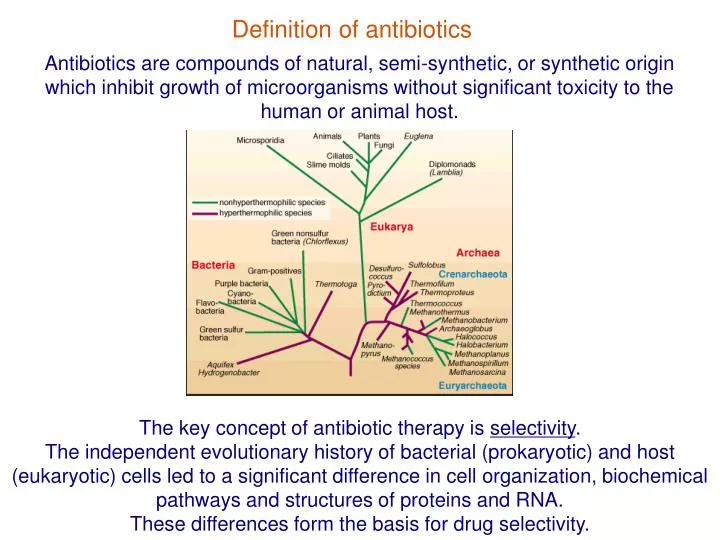
Definition of antibiotics. Antibiotics are compounds of natural, semi-synthetic, or synthetic origin which inhibit growth of microorganisms without significant toxicity to the human or animal host. The key concept of antibiotic therapy is selectivity .

E N D
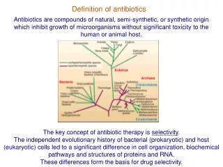
Definition of antibiotics Antibiotics are compounds of natural, semi-synthetic, or synthetic origin which inhibit growth of microorganisms without significant toxicity to the human or animal host. The key concept of antibiotic therapy is selectivity. The independent evolutionary history of bacterial (prokaryotic) and host (eukaryotic) cells led to a significant difference in cell organization, biochemical pathways and structures of proteins and RNA. These differences form the basis for drug selectivity.
Bases of antibiotic selectivity 1. The target of an antibiotic can be present only in bacteria but not in the eukaryotic host. 2. The target in bacteria is different from the homologous target in the eukaryotic host. Modern genomics provide a great tool for identifying targets of new selective antibiotics
Selectivity of antibiotics is not ‘natural’ Natural antibiotics are weapons that bacteria or fungi use to compete with other microorganisms. Selectivity is not a ‘natural’ feature of antibiotics. Most of clinically-useful antibiotics are fortuitously selective antibacterials. Many antibiotics are omni-potent and inhibit growth of a wide variety of organisms. Such antibiotics can be developed into selective drugs through modification of their chemical structures.
Antibiotics are classified as bacteriostatic or bactericidal. Bacteriostatic drugs make bacteria dormant, but do not kill them. Most bacterial cells resume growth after removal of the antibiotic (e.g. chloramphenicol) Bactericidal drugs kill bacteria (e.g. ciprofloxacin) Antibiotics with a bactericidal mode of action are preferred, especially for treatment of immunocompromised patients. The mode (static vs. cidal) of antibiotic action may differ for different pathogens and may depend on the drug concentration. The basis of bactericidal versus bacteriostatic effects is poorly understood but maybe related to the accumulation of reactive oxygen radicals in the bacterial cells upon treatment with bactericidal drugs.
After the golden era of the 1940s-1950s, the progress in antibiotic discovery has significantly slowed down until the year 2000 Golden era in antibiotic discovery No principally new antibiotics 1949 1947 2003 1920 1942 1952 1958 1962 2000 linezolid -lactams daptomycin macrolides tetracycline streptogramins lincosamides sulfonamides glycopeptides aminoglycosides Growing resistance
The appearance and spread of antibiotic resistance calls for new antibiotics. Resistance to the available antibiotics is a result of Darwinian selection There are two major mechanisms by which bacteria can become resistant to antibiotics: 1. Mutation of a “normal” bacterial gene resulting in antibiotic resistance. 2. Acquisition of a resistance gene from the environment. Sensitive bacteria spontaneous mutation in the bacterial gene acquisition of a resistance gene (often brought by a genetic vector) Resistant bacteria Resistant bacteria vertical transfer (to the descendants) horizontal transfer (gene exchange) vertical transfer (to the descendants)
There are three major types of resistance mechanisms: • Modification of the drug (aminoglycoside modifying enzymes) • Modification of the drug target (Erm methylases, target site mutations) • Reducing the drug’s intracellular concentration: drug-specific transporters (mefA), multi-drug transporters (bmr), reduced drug uptake (mutations in porins)
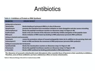
The number of targets of clinically-useful antibiotics is very limited. Cell Wall Biosynthesis Beta-lactams Glycopeptides Bacitracin Protein synthesis Aminoglycosides Oxazolidinones Tetracyclines Macrolides Chloramphenicol Lincosamides Streptogramins Folate Biosynthesis Sulfonamides Membrane Integrity Cationic peptides Lipopeptides DNA Gyrase Fluoroquinolones RNA Polymerase Rifampicin Many resistant strains are cross-resistant to different drugs acting upon the same target.
New antibiotics Originally, screening for new antibiotics was based on testing the bacterial or fungal extracts. A number of drugs developed in the middle of the 20th century still remain among the best. These include aminoglycosides, cephalosporins, tetracyclines, macrolides, etc. There is now a renewed interest in screening natural sources (especially, non-traditional, e.g. marine microorganisms)
New antibiotics: genome-based approaches Genomics holds a good potential for identifying new drug targets.
New antibiotics: Functional genomics approach gene target HTS enzyme inhibitors lead lead optimization drug candidate preclinical clinical In the functional genomics approach, the enzymatic target is first defined and then inhibitors are searched for in high-throughput screening assays (HTS). “easy” hard On average: only one lead candidate is developed from 14 high throughput screening experiments!
New antibiotics: Reverse genomics approach HTS growth inhibitors lead target mode of action lead optimization drug candidate preclinical clinical In the reverse genomics approach the lead is first identified in the whole cell assay and then the target is searched for.
Cell wall as antibiotic target Most of the bacteria have a rigid cell wall which protects the cell from changes in osmotic pressure. Presence of the cell wall is critical for the survival of bacterial cell. The structure and composition of bacterial cell wall is dramatically different from the cell envelope of the eukaryotic cell. Therefore, enzymes of cell wall biosynthesis are unique to bacteria and presents an excellent target for antibiotics. According to the structure of their cell wall, all bacteria are divided into Gram-positive and Gram-negative according to a staining procedure developed by Christian Gram in 1884.
Cell wall of Gram-positive bacteria is thick and unstructured Outside of the cytoplasmic membrane of Gram-positive bacteria lies a thick layer of peptidoglycan which determines the rigidity of the cell wall. In Gram-positive bacteria, peptidoglycan accounts for 50% of the cell weight and up to 90% of the weight of the cell wall. Peptidoglycan layer is 20-80 nm thick and rather unstructured.
Cell wall of Gram-negative bacteria is thin and neatly organized Periplasmic space The cell wall of Gram-negative bacteria consists of the cytoplasmic membrane, a thin layer of peptidoglycan, and an outer membrane. The area between the cytoplasmic membrane and peptidoglycan layer is called the periplasmic space.
Peptidoglycan composition Peptidoglycan consists of polysaccharide chains cross-linked by peptide ‘bridges’. 35% to 50% of peptides attached to polysaccharide chains are crosslinked. Peptide tails protrude from the polysaccharide helically and thus crosslink to different polysaccharide chains. This accounts for the rigidity of the cell wall.
Peptidoglycan biosynthesis The precursor monomers of the peptidoglycan polymer, disaccharide-pentapeptides, are synthesized in the cell cytoplasm, transported across cytoplasmic membrane, and then attached to the growing peptidoglycan polymer.
Transpeptidase D-Ala D-Ala D-Ala Crosslinking between glycan strands is catalyzed by the enzyme called transpeptidase (TPase). In the course of the reaction, TPase hydrolyzes peptide bond between two terminal D-Ala residues of the precursor and forms a transient covalent link with the precursor peptide. The intermediate is then resolved with the formation of a new peptide bond. The reaction of formation of the covalent intermediate is targeted by -lactam antibiotics.
Penicillin was discovered by A. Fleming in 1929 Ernst Chain Howard Florey Alexander Fleming Photo: L. Segovia Development of methods for growing Penicillium notatum and purifying penicillin by Florey and Chain made it into a drug. The deep fermentation method, the use of corn steep liquor and the discovery of P. chrysogenum by Mary Hunt made the commercial production of penicillin possible.
-lactam antibiotics O N R N O S O H 3 carbapenems penicillins cephalosporins monobactams The most important class of antibiotics affecting cell wall biosynthesis are -lactams. -lactam group (a four-atom cyclic amide) is the pharmacophore of all -lactam antibiotics. -lactam rings were unknown before the discovery of penicillin and it took big effort to determine the structure of the drug. The most important classes of -lactam antibiotics are penicillins, cephalosporins, carbapenems and monobactams.
Mechanism of -lactam action CH CH 3 3 The mechanism of action of -lactam antibiotics is based on the similarity of structures of the C - N bond in the -lactam ring and the peptide bond connecting two D-alanine residues of the peptidoglycan precursor. TPase recognizes the -lactam as its substrate and forms a covalent bond with the antibiotic molecule. This adduct is very stable and because of that TPase is irreversibly inactivated. penicillin D-Ala-D-Ala C N C N C COOH H H O Covalent complex of penicillin with TPase
Penicillin binding proteins (PBPs) Several other enzymes of cell wall biosynthesis with a mechanism of action comparable to TPase are also targeted by -lactams. These proteins can be detected on a gel by their ability to bind penicillin. These proteins are called ‘penicillin binding proteins’ or PBPs.
Penicillins : penicillin G In penicillins, the -lactam ring is fused to thiazolidine ring. Originally, penicillin was produced in the form of Penicillin G (benzylpenicillin) by fermenting Penicillium mold in the presence of phenyl acetic acid Good activity, but only against Gram-positive bacteria Acid- and alkali-labile Sensitive to the action of inactivating penicillinases
6-APA R C O N H Presently, many penicillins are produced semisynthetically starting from 6-aminopenicillanic acid (6-APA) as a precursor. 6-APA can be generated from penicillin G by cleaving off the benzyl moiety of penicillin G. Various new side chains can be then attached to the penicillin molecule through the amino group of 6-APA Benzyl-penicillin New penicillins 6-APA CH3 CH3 S S H2N CH3 CH3 N N O O CO2H CO2H Various penicillins differ mainly by the nature of the N6 side chain R
Penicillin improvements: better acid stability C H S 3 C O N H H O C H C H 3 N N H O 2 O H C 2 Amoxicillin Cloxacillin H O C H 3 N C H S 3 C O N H C H 3 Cl N O C O H 2 amide bond The amide bond in the β-lactam ring is highly strained and relatively unstable in acidic solutions. The rate of acid hydrolysis depends on the chemical nature of the side chain. Electron-withdrawing side chains decrease the rate of acid hydrolysis. Because of that, amoxicillin or cloxacillin are more acid-stable: they can withstand the acidic pH of the stomach and can be used orally.
Penicillin improvements: broader spectrum Penicillins enter the periplasmic space of Gram-negative bacteria through the ‘holes’ in the outer membrane (porins). Hydrophobic side chains (e.g. benzyl group in penicillin G) interfere with passage through porins. More polar groups, such as -NH2 or -COOH facilitate crossing the outer membrane and increase access of β-lactmas to the periplasmic space of Gram-negative bacteria. ampicillin C H S 3 CH CONH C H 3 N N H O 2 C O H 2 amoxicillin C H S 3 C O N H H O C H C H 3 N N H O 2 C O H 2 carbenicillin C H S 3 CH CONH C H 3 N COOH O C O H 2
The pro-drug approach can be used to increase bioavailability of some penicillins CH NH2 Ampicillin has a very broad spectrum of activity. It can be used orally or parenterally. But it has low bioavailability. A more lipophilic pro-drug, pivampicillin, has a better oral bioavailability. Pivampicillin is an ester of ampicillin; the ester bond is slowly hydrolyzed in the blood resulting in the release of the active ampicillin. ampicillin C H S 3 CH CONH C H 3 N N H O 2 C O H 2 pivampicillin C H S 3 CONH C H 3 N O CO2CH2OCOC(CH3)3
Penicillin improvements: resistance to -lactamases The main mechanism of resistance to penicillin is based on the secretion by bacteria of enzymes -lactamases that can hydrolyze amide bond in -lactam ring. The presence of a bulky side chain in the drug may hinder access of a -lactamase to the amide bond. Therefore, penicillin derivatives containing bulky side chains are fairly resistant to the -lactamase action. cloxacillin methicillin H O C H 3 O C H 3 N C H C H S 3 S 3 C O N H C O N H C H C H Cl 3 N 3 N O C O H O O C H C O H 2 3 2
-lactamase inhibitors CH2OH O O O S N N O O COOH COOH A useful way to overcome -lactamase-based resistance is to administer a -lactam drug in combination with -lactamase inhibitors. Such inhibitors (clavulanic acid, sulbactam, tazobactam) possess a -lactam ring and generally resemble -lactam antibiotics. They function by binding to the -lactamase enzymes and inactivating the enzyme without being degraded. Such inhibitors, which look like -lactam antibiotics, have only weak antimicrobial activity. Popular combinations are amoxicillin with clavulanate (augmentin) or ampicillin with sulbactam (unasyn). Clavulanic acid Sulbactam
Other mechanisms of resistance to -lactams An important mechanism of resistance to -lactams involves mutations in transpeptidases and other penicillin-binding proteins (PBPs) involved in bacterial cell wall biosynthesis. Resistance mechanism found in methicillin-resistant Staphylococcus aureus (MRSA) is based on acquisition of a mecA gene which encodes a resistant mutant protein, PBP2’. PBP2’ has a very low affinity for -lactam antibiotics and can support cell wall biosynthesis even when all other PBPs are covalently inactivated by the drug. Genetic analysis show that mecA gene has been independently transferred to S. aureus at least 5 times resulting in 5 independent lineages of MRSA
Cephalosporins Cephalosporins have been first obtained from a fungus Cephalosporium acremonium. Similar to penicillins, many cephalosporins are produced semi-synthetically either starting from 7-aminocephalosporanic acid (7-ACA) or by converting relevant penicillins into cephalosporins. 7-aminocephalosporanic acid (7-ACA) S 7 H2N 3 N O C H 3 O 2 C O H O 2 The activity of cephalosporins is modulated not only by the nature of substitutions R2 at C7 (as in penicillins) but also by the side chain R1 at C3.
Cephalosporins are classified by generations N H 2 N H S S O C H O 2 N H C O N H C O S S S N O C H N N O C H N H C O N H C O 3 N N O C H O 3 3 N C H S N Cl H C O C O H O N 2 S 2 N O 2 N O N H C O C O H H C C O H H N 2 3 N 2 C H O C O C H 2 S 2 3 O S C O H N N H C O 2 N O C H C O H N N S N S C H 2 2 N 3 O S N N N C O H N H C O 2 H N N 2 S C H O 2 C O H 2 cephalothin cafazolin cephalexin I Active against Gram-positive cocci and streptococci. cefamandole nofate cephaclor II Improved activity against some Gram-negatives, for example, H. influenzae. cefotaxime cefixime III Better activity for Gram-negatives though, somewhat reduced activity against Gram-positive pathogens.
Moxalactam C O O H O C H 3 S C H 3 N H C O 7 N N S N O H O N N C O H 2 Similar to penicillins, cephalosporins can be inactivated by -lactamases. Resistance to -lactamases increases in drugs such as moxalactam which have bulky side chains. The presence of 7--methoxy group increases moxalactam resistance to -lactamase hydrolysis even further.
Carbapenems OH H H H S NH2 H3C N O CO2H Carbapenems combine chemical features of penicillins and cephalosporins. Prototype carbapenem thienamycin, has been isolated from Streptomyces cattleva. It exhibits excellent activity against a broad spectrum of Gram-positive and Gram-negative organisms. thienamycin Thienamycin penetrates very easily through the outer membrane of Gram-negative bacteria (through porin "holes") . It is resistant to the action of extended spectrum -lactamases (ESBL) which can inactivate penicillins and cephalosporins. In contrast to penicillins and cephalosporins which target only PBPs, carbapenems can target another enzyme of the cell wall biosynthesis, Ld transferase (LdT) which sometimes can help cell to bypass the need for TPase. Therefore, carbapenems shows excellent activity against some Gram-positive strains which developed resistance to penicillins and cephalosporins.
Imipenem OH H H H S NH2 OH H H H3C N H S O CO2H N H N H H3C N O CO2H imipenem thienamycin In concentrated solutions, the side amino group of thienamycin can react with the amide bond in the -lactam ring of another thienamycin molecule making the drug unstable in concentrated solutions. This problem has been solved in the thienamycin derivative, imipenem by modifying the side chain. Imipenem was the first parenteral carbapenem. doripenem ertapenem meropenem The newer drugs of this class are meropenem, ertapenem and doripenem The drawback of carbapenems is that they are acid-labile and therefore used only intravenously. In addition, they are very expensive.
Monobactams Aztreonam (Azactam) Monobactams were developed as narrow-spectrum antibiotics specifically targeting aerobic Gram-negative bacteria. Monobactams are particularly useful for the treatment of individuals allergic to penicillin. Such patients can still be treated with the monobactams, which are sufficiently structurally different to not induce allergic reaction.
Cell wall inhibitors of non--lactam type: Bacitracin A number of peptide antibiotics affect cell wall biosynthesis. Bacitracin, a polypeptide antibiotic produced by licheniformis group of Bacillus subtilis, inhibits cell wall synthesis by interfering with dephosphorylation of the lipid carrier that moves the peptidoglycan precursors across the cytoplasmic membrane. Blocking regeneration of the lipid carrier aborts cell wall synthesis. Side effect of bacitracin is based on its interference with sterol biosynthesis in mammalian cells which accounts for its certain toxicity in humans. Therefore, it is used exclusively in topical formulations.
Cell wall inhibitors of non--lactam type: Vancomycin An important class of antibiotics that interfere with synthesis of cell wall are glycopeptides. The most important of them is vancomycin. Vancomycin is isolated from a bacterium Nocardia orientalis. Vancomycin is a tricyclic glycopeptide with a large molecular weight of 1449 D. It is active against most Gram-positive bacteria. It is especially important in the treatment of infections due to methicillin- and cephalosporin-resistant organisms. Vancomycin is bactericidal against most of the susceptible bacteria.
Vancomycin vancomycin D-Ala-D-Ala Vancomycin binds very tightly to the D-Ala - D-Ala residues at the ‘end’ of the peptidoglycan precursor peptide. Because of that, the peptide bond between two D-Ala residues becomes inaccessible to TPase so that peptidoglycan strands cannot be crosslinked.
Resistance to vancomycin Vancomycin was kept as a ‘reserve’ antibiotic and was usually prescribed only when other drugs proved to be inactive. However, even in spite of its relatively infrequent usage, resistant strains eventually appeared. Vancomycin resistant enterococci (VRE) account now for up to 25% hospital strains of enterococci. First vancomycin resistance appeared in staphylococci in ‘vancomycin intermediate susceptible S. aureus’ - VISA. VISA cells have abnormal peptidoglycan: the cell wall is thicker and less crosslinked. Therefore more D-Ala - D-Ala remain in the cell wall and are exposed. They work as vancomycin trap (sponge). However, because of the abnormal cell wall, VISA strains are sick (high fitness cost) and do not spread very rapidly. In 2002 the first Vancomycin-resistant S. aureus (VRSA) strain was reported. The Van gene they have acquired replaces D-Ala - D-Ala in the precursor for D-Ala - D-lactate. Since Van genes are active only when cells are exposed to vancomycin, VRSA strains are more fit than VISA.
New glycopeptides Newer versions of glycopeptide antibiotics include teicoplanin and dalbavancin (ZevenTM). These drugs are similar to vancomycin but have additional hydrophobic side chains. Teicoplanin and dalbavancin are active against all vancomycin-sensitive strains, plus against a number of resistant strains and do not induce expression of VanB resistance gene. Both teicoplanin and dalbavancin are rapidly cidal. Dalbavancin is very tightly bound to serum proteins and is therefore very stable. Because of that, it can be administered once a week. dalbavancin