Download
1 / 58
1.11k likes | 4.94k Views
PEPTIC ULCER disease (PUD) Pathophysiology. By Dr. Abdelaty Shawky Assistant professor of pathology. * Anatomy of the stomach: The stomach is located in the upper part of the abdomen just beneath the diaphragm .

E N D
PEPTIC ULCER disease (PUD)Pathophysiology By Dr. Abdelaty Shawky Assistant professor of pathology
* Anatomy of the stomach: • The stomach is located in the upper part of the abdomen just beneath the diaphragm . • The stomach is distensible and on a free mesentery, therefore, the size, shape, and position may vary with posture and content.
An empty stomach is roughly the size of an open hand and when distended with food, can fill much of the upper abdomen and may descend into the lower abdomen on standing.
The stomach may be divided into seven major sections. The cardia is a 1–2 cm segment distal to the esophagogastric junction. • The fundus refers to the superior portion of the stomach that lies above an imaginary horizontal plane that passes through the esophagogastric junction.
The antrum is the smaller distal one-fourth to one-third of the stomach. The narrow 1–2 cm channel that connects the stomach and duodenum is the pylorus. • The lesser curve refers to the medial shorter border of the stomach, whereas the opposite surface is the greater curve.
Histology of stomach • Mucosa. • Submucosa (with Meissner plexus). • Muscularis propria (outer longitudinal layer, Auerbach/myenteric plexus, inner circular layer, innermost oblique layer). • Serosa.
Gastric mucosa has two compartments:1. Superficial foveolar compartment, relatively uniform throughout, consists of surface epithelial / foveolar cells lining the entire mucosal surface and pits2. Deep glandular compartment, varying composition and thickness in different regions containing gastric glands
The gastric mucosa is very metabolically active • The entire mucosa is replaced every 2 to 8 days. • Mucous neck cells at the base of foveola are progenitors of surface epithelium and gastric glands, mitotically active, site of gastric carcinogenesis. • Ratio of foveola to gland volume differs by region: cardia/antrum: 50/50; fundus: 25/75
Cardia Branching mucous glands without parietal cells.
Antrum Branching mucus glands, cytoplasm may be bubbly, vacuolated, granular or glassy
Fundus • Straight glands composed of tightly packed chief cells, parietal cells, endocrine cells, mucus cells • Higher ratio of glands to foveola than antrum
Parietal cells • Primarily in fundus/body. • Have eosinophilic cytoplasm. • Produce acid via H+/K+ ATPase pump. • Also secrete intrinsic factor which binds luminal Vitamin B12. • Stimulated by vagus nerve, binding of gastrin receptor by gastrin from antral cells, binding of H2 receptor by histamine from enterochromaffin-like cells.
Mucous cells • Produce neutral mucin. • Lightly eosinophilic or clear cytoplasm and bubbly
Chief cells • Scattered in fundus/body. • Have basophilic cytoplasm. • Release pepsinogen I and II, which are activated by low luminal pH to pepsin
Endocrine cells • Scattered in fundus/body. • Have clear cytoplasm. • Produce: • Histamine (enterochromaffin-like). • Gastrin (G cells). • Serotonin (enterochromaffin cells). • Somatostatin (D cells).
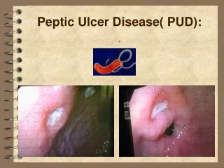
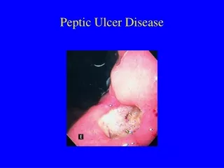
* Definition: Ulceration of any part of the G.I.T exposed to the gastric juice.
Acute peptic ulcers (stress ulcers)
* Etiology: I. Infective: Salmonellosis & staph. Food poisoning. II. Non infective: • Drugs; NSAI, cytotoxic drugs and cortisone. • Alcoholism. • Cigarette smoking. • Uremia. • Severe stress. • Chemical irritation: by strong alkalis, acids. • Mechanical trauma; during endoscopic examination.
* Sites:stomach & 1st part of duodenum. * Pathology: hemorrhagic inflammation and superficial ulceration. * Fate: usually heal by regeneration.
* Sites of peptic ulcer: • Duodenum: First portion usually toward the anterior wall is more often affected. • Stomach: Usually antrum toward the Lesser curvature. • At the margins of a gastroenterostomy (stomal ulcer) • In the duodenum, stomach or jejunum of patients with Zollinger-Ellison syndrome. • Within Meckel’s diverticulum that contains ectopic gastric mucosa.
* Etiology: • Genetic factors:Genetic factors play a role in the pathogenesis of ulcer disease. The lifetime prevalence of developing ulcer disease in first-degree relatives of ulcer patients is about three times greater than the general population. Approximately 20–50% of duodenal ulcer patients report a positive family history.
2. Hormonal factors: evident in in cases of Zollinger Ellison syndrome. 3. Environmental factors: cigarette smoking, coffee and alcoholism. 4. Emotional stress: evident with duodenal ulcer. 5. Associated diseases: Chronic bronchitis, emphysema, liver cirrhosis, renal failure.
* Pathogenesis: 1. Duodenal ulcer: • Due to increased gastric juice secretion + Rapid emptying of stomach into duodenum * Causes: • Increased vagus nerve stimulation. • Increased number of parietal cell (as in tall thin patient). • Increase hormonal stimulation (Z.E syndrome) Gastrinoma Gastrin. • Increase the responsiveness of parietal cells to secretory stimuli. • psychosomatic stimuli.
2. Gastric ulcer. • Due to decreased mucosal defense mechanisms or increasing damaging forces…
* Mucosal defense mechanisms: • Bicarbonate secretion • Mucous secretion • Tight adherence between epithelial cells to prevent any acid leakage to the inside. • Good blood supply to the mucosa • Renewal of damaged epithelial cells.
* Damaging agents: • H. pylori infection. • Increased gastric acid secretion. • Drugs: NSAI, Aspirin, cortisone and cytotoxic drugs.. • Cigarette smoking. • Chronic alcoholism.
H. Pylori infection • H. pylori is the etiologic factor in most patients with peptic ulcer disease and may predispose individuals to the development of gastric carcinoma. H. pylori colonizes in the human stomach.
The method of H. pylori transmission is unclear, but seems to be person-to-person spread via a fecal-oral route. The prevalence of H. pylori in adults appears to be inversely related to the socioeconomic status. It is also thought that water is a reservoir for transmission of H. pylori.
H. pylori infection is present in almost all patients with duodenal ulcers and 70% of cases with gastric ulcers. * Mechanism: 1. H. pylori secretes urease (generates ammonia), protease (breaks down glycoprotein in the gastric mucus) and phospholipases.
2. Bacterial lipopolysaccharide attracts inflammatory cells to the mucosa. Chronically inflamed mucosa are susceptible to injury. 3. A bacterial platelet-activating factor promotes thrombotic occlusion of surface capillaries.
Non-steroidal Anti-Inflammatory Drugs (NSAIDS) • Risk factors for the development of NSAID-associated gastric and duodenal ulcers include advanced age, history of previous ulcer disease, higher doses of NSAIDs. • NSAIDs initiate mucosal injury topically by their acidic properties.
Systemic effects of NSAIDs appear to play a predominant role through the decreased synthesis of mucosal prostaglandins. The precursor of prostaglandins, arachidonic acid, is catalyzed by the two cyclo-oxygenase isoenzymes, cyclo-oxygenase-1 and cyclo-oxygenase-2.
Literature suggests that the anti-inflammatory properties of NSAIDs are mediated through inhibition of cyclo-oxgenase-2, and adverse effects, such as gastric and duodenal ulceration, occur as a result of effects on the constitutively expressed cyclo-oxygenase-1.
Zollinger-Ellison Syndrome • The classic triad of Zollinger-Ellison syndrome involves: • Peptic ulcers in unusual locations (the jejunum) in addition to duodenal and gastric sites. • Massive gastric acid hypersecretion. • Gastrin-producing islet cell tumor of the pancreas (gastrinoma).
* Gross features: • Site:Gastric ulcers are located at the antrum toward the lesser curvature. The duodenal ulcer is usually located at the 1st part anteriorly. • Shape:Round, oval. • Size:Usually less than 4 cm in diameter.
Depth of the ulcer: - Superficial ulcer penetrate the mucosa reaching up to the muscularis mucosa. - Deeply excavated ulcers having their bases on the muscularispropria.
When the entire wall is penetrated, the base of the ulcer may be formed by adherent pancreas, omental fat, or liver. Free perforation into the peritoneal cavity may occur.
Edge: - Sharp edge. • Floor: • Clean • Base of ulcer: - Firm (formed of bundles of muscles and fibrous tissue). • Margin (Surrounding gastric mucosa): - Edematous and reddened due to gastritis.
Biopsy of peptic ulcer • Biopsy is necessary to distinguish between benign and malignant ulcers. • Biopsy should be taken from the ulcer edge, at least from each quadrant. • Up to 10-12 biopsies may be taken to exclude cancer.
* Microscopic features: • Active peptic ulcer shows; • Superficial zone:fibrinoid necrosis & neutrophils. • Intermediate zone:chronic inflammatory cells & granulation tissue. • Deep zone:fibrous tissue, muscle remnants.