Download
1 / 1
10 likes | 156 Views
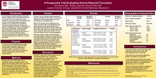
A Prospective Trial Evaluating Scleral Rebound Tonometry Sara Duke , MD, Andrew Logeman, Shuchi Patel, MD Loyola University Chicago, Department of Ophthalmology, Maywood IL. Introduction. Results. Results. Demographics/Characteristics.

E N D
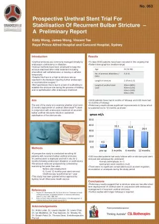
A Prospective Trial Evaluating Scleral Rebound Tonometry Sara Duke, MD, Andrew Logeman, Shuchi Patel, MD Loyola University Chicago, Department of Ophthalmology, Maywood IL Introduction Results Results Demographics/Characteristics Current devices used to estimate intraocular pressure (IOP) perform the measurements on the cornea. This is not possible in some patients such as those with a keratoprosthesis or other corneal pathology. Glaucoma is known to occur in about 75% of patients following a keratoprosthesis, but accurate pressure readings to monitor for adequate IOP control are not possible. Previous studies have examined the use of the TonoPen, Schiotz tonometer and pneumotonometer on the sclera to measure IOP with limited success, and none have repeatedly shown a statistically significant correlation to corneal tonometry. Patients included (analyzed) 101 (59) Eyes included (analyzed) 184 (116) Male:Female 28:31 Mean age (range) 65 (37-90) Mean spherical equivalent -0.21 -7.00 to-2.01 15.7% -2.00 to +2.00 78.5% +2.01 to +3.25 5.8% Past Ocular History (eyes) Glaucoma 39 Glaucoma suspect 39 Ocular hypertension 3 Pseudophakic 27 Diabetes 14 Thyroid eye disease 2 184 eyes from 101 patients ages 24-90 years old were included in this study. 116 eyes from 59 patients were analyzed as 68 eyes were omitted due to incomplete data. Mean GAT IOP was 15.91 mmHg (SD 4.13), mean corneal RT was 14.50 mmHg (SD 4.24) and mean scleral RT was 48.84 (SD 21.41). Mean spherical equivalent refraction (SE) was -0.21 D (SD 2.05), mean CCT was 547.68 µm (SD 45.65), mean AL was 24.06 mm (SD 1.21). 89 eyes were phakic and 27 were pseudophakic. Pearson analysis reveals a strong positive correlation between GAT and corneal RT (0.77) but weak positive correlation between GAT and scleral RT (0.22) as well as corneal RT and scleral RT (0.22). This trend persists when phakic and pseudophakic eyes are evaluated independently. Multivariate regression analysis was also performed and did not find a correlation between scleral RT and GAT when accounting for GAT, CCT, AL and SE. Average Standard (mmHg) Deviation GAT 15.91 4.13 Corneal RT 14.50 4.24 Scleral RT 48.84 21.41 Average Standard Deviation CCT (µm) 547.68 45.65 AL (mm) 24.06 1.21 SE (D) -0.21 2.05 Purpose To determine if a predictable relationship exists between the gold standard of IOP measurement, Goldmannapplanation tonometry (GAT), and scleral rebound tonometry (RT) to provide an accurate and reliable assessment of IOP via scleral measurements. Conclusions • Scleral RT shows consistently higher IOP measurements than the corneal IOP measurements from the same eye. • Scleral RT has a statistically significant positive correlation to corneal RT. • In this study, scleral RT does not provide accurate and reliable IOP measurements when compared to GAT even after adjusting for phakic status, CCT, AL and SE. • Further studies are needed to establish an accurate and reliable method of scleral tonometry such that patients with a keratoprosthesis and glaucoma can be better monitored. Discussion Scleral RT shows IOP measurements that are consistently higher than corneal IOP measurements by roughly 33-34 mmHg. Corneal RT correlates well to GAT; unfortunately, scleral RT measurements have poor correlation to corneal measurements independent of phakic status. Analysis of scleral RT with relation to GAT, CCT, AL and SE concurrently fails to reveal a statistically significant regression model in either phakic or pseudophakic eyes. This is likely due to the increased number of variables which effect scleral rigidity. In addition, the overlying conjunctiva undoubtedly contributes to inconsistent scleral IOP readings. Multivariate regression model assessing the relationship between scleral rebound tonometry and ophthalmic variables as listed above. Methods A prospective non-randomized trial of individuals 18 years of age and older. Each had his/her IOP measured by GAT, next corneal RT then scleral RT on the inferotemporal sclera. The patient’s age, gender, refractive error, central corneal thickness (CCT), axial length (AL) and phakic status were recorded. Pearson’s correlation and multivariate regression analysis were used for statistical examination. References • Barraza RA, Sit AJ. Investigation to determine a relationship between scleral and corneal tonometry. ARVO poster presentation; May 02, 2010. • Brusini P, Salvetat ML, Zeppieri M, Tosoni C, Parisi L. Comparison of ICare tonometer with Goldmannapplanation tonometer in glaucoma patients. J Glaucoma 2006; 15(3): 213-217. • Erlich JR, Haseltine S, Shimmyo M, Radcliffe NM. Evaluation of agreement between intraocular pressure measurements using Goldmannapplanation tonometry and Goldmann correlated intraocular pressure by Reichert’s ocular response analyser. Eye 2010; 24(10); 1555-1560. • Duke SL, Abugo U, Patel S. A prospective trial comparing scleral pneumotonometry to Goldmannapplanation tonometry. ARVO poster presentation; May 10, 2012. • Shen CC, Downs J, Mansberger SL. Assessment of intraocular pressure along the limbus and sclera using contact and indentation tonometry. ARVO poster presentation; May 02, 2010. Acknowledgements: This work was supported by the Richard A. Perritt Charitable Foundation.