Scrotal Swelling: Diagnosis and Management Overview
670 likes | 1.12k Views
Learn about the anatomy, diagnosis, and differential diagnosis of scrotal swelling. Understand the approach and management of painful and painless scrotal conditions. Detailed examination and investigation guidelines provided.

Scrotal Swelling: Diagnosis and Management Overview
E N D
Presentation Transcript
Scrotal Swelling RawanAlshabeeb AfnanAlmarshadi Supervised by: Dr. Hamdan Al- Hazmi
Outline • Anatomy of the scrotum • Differential diagnosis • Approach to a patient with scrotal swelling • Painfull scrotal swelling • Painless scrotal swelling
The wall of scrotum has the following layers(imp for mcq) 1-skin 2-superficial fascia 3-external spermatic fascia derived from the external oblique 4-cremasteric muscle derived from the internal oblique 5- internal spermatic fascia derived from the fascia transversalis 6-tunica vaginalis(remnant of Peritoneum )
Coverings of the spermatic cord:* Tunica vaginaliscovers the anterior surface of the spermatic cord just above the testis* Internal spermatic fascia (transversalis/endoabdominal fascia)* Cremasteric fascia (fascia of internal oblique muscle)* External spermatic fascia (aponeurosis of the external oblique muscle)* The cremasteric fascia contains loops of cremasteric muscle, which draws the testis superiorly in the scrotum when it is cold.
Contents of spermatic cord • Ductus deferens (conveys sperm from the epididymis to the ejaculatory duct)* Arteries* Testicular artery (arises from the abdominal aorta at L2)* Artery of the ductus deferens (arises from inferior vesical artery)* Cremasteric artery (arises from the inferior epigastric artery)* Veins* Pampiniform plexus (formed by up to 12 veins, drain into right and left testicular veins)* Nerves* Sympathetic nerve fibers on arteries* Sympathetic and parasympathetic nerve fibers on the ductus deferens* Genital branch of the genitofemoral nerve supplying the cremaster muscle* Lymphatics* Lymphatic vessels draining the testis and closely associated structures* lumbar lymph nodes
In the acute scrotum our main goal is to detect or exclude a testicular torsion
We have We have 3 ways of DDX must say them all in exam ; 1- acute vs chronic 2- painful vs painless 3- get above it vs can’t
History • timing of onset: acute or insidious onset • associated symptoms or prior episodes • age at presentation • Physical examination • general appearance • lie of testes(to diffrentiate between torsion and epidiymo orchitis), scrotal skin, fluid collection, • testes or epididymis tenderness • Get above the swelling ?
Investigation: • Urinalysis: bacteria, WBC’s, crystals • commonly in epididymitis • Obtain urine culture(why ? If pt have +ve culture with epidedmytise R/O congenital anomaly by US or MCUG (in pediatrics ) • CBC may be helpful • Radiographic studies • Ultrasonography , Nuclear Scan • Doppler US.
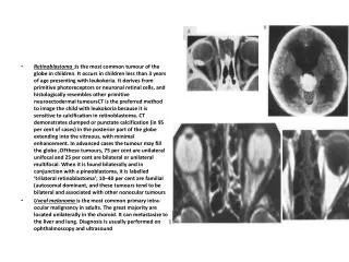
Diagnostic test Color Doppler ultrasound FIGURE 1. Color Doppler ultrasonogram showing acute torsion affecting the left testis in a 14-year-old boy who had acute pain for four hours. Note decreased blood flow in the left testis compared with the right testis • Noninvasive assessment of anatomy and determining the presence or absence of blood flow. • sensitivity: 88.9% specificity of 98.8% • operator dependent. • .
Color Doppler ultrasound FIGURE 2. Color Doppler ultrasonogram showing late torsion affecting the right testis in a 16-year-old boy who had pain for 24 hours. Note increased blood flow around the right testis but absence of flow within the substance of the testis. FIGURE 3. Color Doppler ultrasonogram showing inflammation (epididymitis) in a 16-year-old boy who had pain in the left testis for 24 hours. Note increased blood flow in and around the left testis.
Color Dopplar US is imp to differentiate between epidedmytis and torsion , the first we will see high blood supply in the affected site(infection) while in the second decrease blood supply(torsion )
1-Testicular torsion(imp) • It is an Emergency. • Due to twisting of the testis with interference to the arterial blood supply. • May have torsion of cord or appendages. • Incidence is highest between 10-20 y.o.
Clinical Feature • Testicular pain &swelling( Sudden) radiating to the lower abdomen • Nausea and vomiting • previous similar episode • No voiding complaints
Most cases spontaneous torsion. • Anterior surface of each testis run towards the midline.
Types: • Extravaginal: exclusive to perinatal (torsion, the testis, spermatic cord and tunica vaginalis twist en bloc) .It is usually ASYMPTOMATIC(cuz we discover it early before appearnce of symptoms )...and therefore could be managed by observation. • Intravaginal: 90% of adolescent age group. A) extravaginal; (B) intravaginal
- extravaginal in neonates , and means the whole unit torte . • Intravaginalis in adults , means the testes only tort around it self while the tunica vaginalis is not Regarding Rx; • In adults we do a testicular incision • in children we do inguinal incision ? Cuz it’s usually associated with hernia
On Ex: • Swollen, painful, testis drawn up to the groin. • Absent of cremastic reflex on the affected site • Elevation of scrotum doesn’t provide relife of pain (-veprehn sign )
If you in doubt in case of acute painful scrotum so the scrotum must be explored. • If untreated infarction of testis will result. • Untwisting should be carried on within 6 hrs. of symptoms.
The best "test" to diagnose torsion is SURGICAL EXPLORATION once suspected
management • Rx: EMERGANCY • Explore the testis. • Untwist the testis. • If viable so fix to scrotum by anchoring it to scrotal septum and if the other testis is abnormal fix it. • If infracted so remove it.
2-Torsion of testicular appendage(imp): • Most common structure to twist is the appendix of the testis (pedunculated hydatid of morgagni ) • Usually a more gradual onset, pain moderately severe • Blue dot sign. • Age:12 – 24 years age . Blue dot sign.
Management • If dx is in question, surgical exploration Rx ; • If ur not sure if it’s 1 or 2 do an exploration surgery . • If ur sure Rx conservatively • immediate operation with ligation and amputation of the twisted appendage. • when the appendix torsion is late in presentation, it could resemble testicular torsion
3-Testicular trauma • Usually in sports injuries or violance. • may result in bleeding into the layers of tunica vaginalis resulting in haematocele. • S&S: severe pain, scrotal swelling, bruising, tender, enlarged testis.
Management • Investigation: • scrotal ultrasound (beware of an underlying malignancy). • Treatment: CONSERVATIVE • Bed rest • Scrotal elevation • Surgical exploration may needed if: • 1- expanding scrotal hematoma 2- To evcuate the haematocele and to repair the split in tunica albugenea. 3- very sever pain
4- Infections of testis & epididymis • May be acute or chronic. • Acute or chronic orchitis may be due to mumps. • Acute epididymo-orchitis may be due to coliform organisms or gonorrhoea. • Also can follow instrumentation or operations on prostate. • Chronic epididymo-orchitis :common cause of is a partially treated acute one & TB or brucellosis .
clinical features : • pain, edematous, swelling redness of the scrotum, often associated with pyrexia. • +/- symptoms of UTI • In children differentiation from torsion is often impossible and scrotum should be explored. • Enlarged tender testis and epididymis. Prehn sign is +ve Bilatral swelling and pain could be caused by lymphoma
-ve Prehn's sign indicates no pain relief with lifting the affected testicle, which points towards testicular torsion which is a surgical emergency and must be relieved within 6 hours. • Positive Prehn's sign indicates there is pain relief with lifting the affected testicle, which points towards epididymitis.
Management • Investigation: • FBC, MSU, Early morning urine specimens for TB culture. • Treatment: • Acute: Bed rest, Analgesia, • ABx: I.V ciprofluxacin until culture and sensitivity. • Examine the pt in 3 days, if better continue antibiotics, , if pain worsens, consider chronic causes • Chronic: TB-antituberculous drugs. • Orchidectomy if fails. • Long ABx treatment for non tuberculousepididymo-orchitis.
1- Hydrocele; • Is collection of abnormal quantity of serous fluid in the tunica vaginalis.If it contains pus or blood it is called pyocele or haematocele respectively.Hydrocele is more common than the two other varieties.
etiology 1-primary;(newborns) • The cause is unknown • Associated with patency of proccessus vaginalis. • It classified as follows;
1-communicating; it connect with the peritoneal cavity. 2-noncommunicating; it dose not connect with peritoneal cavity.
2- secondary; where the fluid accumulate secondary to pathology inside the testis like epididymo-orchitis,testicular tumor and trauma. infection --- increase production +decrease excretion
Clinical presentation; • Age; • primary hyrocele are most common newborns • Secondary are more common between 20 to 40 years. • Symptoms; • 1-painless swelling 2-frequent and painful micturation may occur if hydrocele is secondary to epididymo-orchitis • Hydrocele not affect fertility
Clinical picture • Examination; • Position; the swelling usually unilateral but can be bilateral .if communicating can not feel the cord above the lump. • Colour and temperature; normal • Tenderness; primary are not tender but secondary may be tender • Composition; fluctuant and have fluid thrill if large enough • Reducibility; can not reduced • Testis impalpable(In communicating type) and transillumenate
Mangement; • Primary; in children • Communicating; • most neonatal hydrocele resolve in first 2 year of life if persists repair as herniotomy(inguinal incision ). NEVER do surgery before 2 years of age.(EXCEPT in 1- very large amount -2- if can’t differentiate between it and hernia 3- increase intrabdominal pressure) NEVER do needle aspiration EVEN in the non- communicating type(cuz it will reaccumulate) • Noncommunicating; • usually resolves spontaneously
In adult; surgical excision; opening the tunica vaginalis longitudinally (scrotal incision ), emptying the hydrocele, everting the sac after excising the redundant sac and suturing the sac behind the cord thus obliterating the potential space • Secondary treatment of the underlying condition Case ; 40 y old man came with painless , transeluminate hydrocele . What's ur next step ? A; do an US for scrotom to R/O testicular tumor
2- Indirect inguinal hernia: • most common ( young , Rt. Side ) • 10% bilateral . • Hernia in babies are a result of persistent processus vaginalis. • If strangulated >> painful and may cause testicular atrophy • Surgery is usually recommended .
Definition • Is dilatation and tortuosity of the pampiniform plexus, which is the network of veins that drain the testicle. • Due to defective valve or compression of the vein by a nearby structure, can cause dilatation of the veins • Very common about 20-30% of normal population will have some degree of varicocele. • More common on left side in 98% of cases. • Bilatral in up to 50% of cases. • Always remember it’s not painful ..
IMP Primary varicocele : is ONLY +ve at standing Secondary varicocele : is when varicocele is +ve at BOTH standing and supine positions. Secondary varicocele could be a sign of a retroperitoneal mass like Renal Cell Carcinoma, Wilms tumor and phaeochromocytoma • Do retroperitonial US to role out renal ca in 2 cases ; 1- varicocele on the rt side 2- secondary .