The Reflex Arc
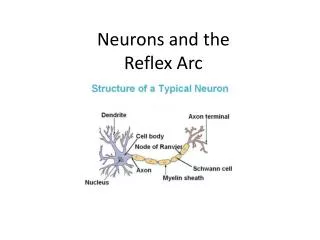
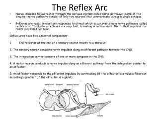
The Reflex Arc. Nerve impulses follow routes through the nervous system called nerve pathways. Some of the simplest nerve pathways consist of only two neurons that communicate across a single synapse .

The Reflex Arc
E N D
Presentation Transcript
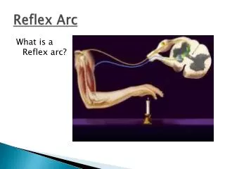
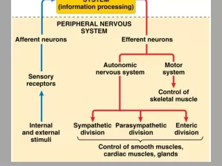
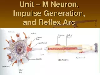
TheReflexArc • Nerve impulses follow routes through the nervous system called nerve pathways. Some of the simplest nerve pathways consist of only two neurons that communicate across a single synapse. • Reflexes are rapid, involuntary responses to stimuli which occur over simple nerve pathways called reflex arcs. Involuntary reflexes are very fast, traveling in milliseconds. The fastest impulses can reach 320 miles per hour. Reflex arcs have five essential components: • The receptor at the end of a sensory neuron reacts to a stimulus. 2. The sensory neuron conducts nerve impulses along an afferent pathway towards the CNS. 3. The integration center consists of one or more synapses in the CNS. 4. A motor neuron conducts a nerve impulse along an efferent pathway from the integration center to aneffector. 5. An effector responds to the efferent impulses by contracting (if the effector is a muscle fiber) or secreting a product (if the effector is a gland).
Patellar (knee-jerk) Reflex • Have your partner sit in a chair or on the edge of a table with legs crossed at the knee. • Find the patella with your fingers • Gently but firmly tap the patellar tendon in the shallow depression immediately below the patella • Compare responses in the right and left knees. 4. Record yourobservations • Diagram and label the reflex arc in your notes. The reflex arc should have labels for the: stimulus, receptor, sensory neuron, spinal cord, motor neuron, and effector. 6. Repeat the above, but have your partner perform Jendrassik's maneuver; that is, clasping his or her hands at chest level in front of your body with fingers locked 7. Have him or her try vigorously to pull the hands apart while you tap the patellar tendon as before.
Ciliospinalreflex • The ciliospinal reflex is an autonomic reflex that can be used to assess multiple conditions, such as deterioration of vital brain tissue and severe head trauma. • The ciliospinal reflex can be elicited by pinching the skin on the nape of the neck while watching the pupils. • The pinch should hurt a bit, but it should not be injurious. The skin on the neck contains the receptors. The smooth muscle of the iris istheeffector. 4. Observe the pupils of both eyes and record the response. 5. Explaintheresults
AccommodationReflex • Focus both eyes on the tip of a pencil held at eye level and at arm's length. • While focusing on the pencil tip, indicate whether or not the objects in the background are in focus. • Keeping the same line of sight, focus on the objects in the background. • What happens to the image of the pencil tip? • During this test you should be looking at the eyes of the person doing the test. • Observe the pupils of both eyes and record the response. • Explaintheresults
Pupillary response • Dim the lights in a room. • After a minute, look at the eyes of another person and note the size of the pupil. • Turn the room lights back on. • Check the size of the pupils again. • Record and explain your results