Download
1 / 32
320 likes | 490 Views
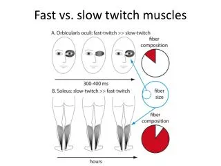
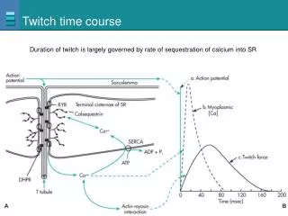
Twitch time course. Duration of twitch is largely governed by rate of sequestration of calcium into SR. X-ray crystal structures.

E N D
Twitch time course Duration of twitch is largely governed by rate of sequestration of calcium into SR
X-ray crystal structures In the beginning of the movie, the myosin heads are in the prestroke ADP-Pi state (yellow) and the catalytic cores bind weakly to actin. Once a head docks properly onto an actin subunit (green), phosphate (Pi) is released from the active site. Phosphate release increases the affinity of the myosin head for actin and swings the converter/lever arm to the poststroke, ADP state (transition from yellow to red). The swing of the lever arm moves the actin filament by ~100 Å; the exact distance may vary from cycle to cycle depending upon the initial prestroke binding configuration of the myosin on actin. After completing the stroke, ADP dissociates and ATP binds to the empty active site, which causes the catalytic core to detach from actin. The lever arm then recocks back to its prestroke state (transition from red to yellow).
Phosphocreatine Provides ATP at beginning of exercise needed for contraction Figure 12-13
Muscle Fatigue Locations and possible causes of muscle fatigue Figure 12-14
Muscle Fiber Types Fast-twitch glycolytic and slow-twitch oxidative muscle fibers Figure 12-15
Length-Tension Relationships in Contracting Muscle Figure 12-16
Summation of Contractions Figure 12-17a
Summation of Contractions Figure 12-17b
Summation of Contractions Figure 12-17d
Motor Units Figure 12-18
Isotonic and Isometric Contractions Figure 12-19
Series Elastic Elements in Muscle Figure 12-20
Muscle Contraction Duration of muscle contraction of the three types of muscle Figure 12-24
Types of Smooth Muscle Figure 12-25a
Types of Smooth Muscle Figure 12-25b
Smooth Muscle • Has longer actin and myosin filaments • Myosin ATPase activity much slower • Actin more plentiful • Has less sarcoplasmic reticulum • IP3-receptor channel is the primary calcium channel
Anatomy of Smooth Muscle Figure 12-27a–b
Smooth Muscle Contraction ECF Ca2+ Sarcoplasmic reticulum 1 Intracellular Ca2+ concentrations increase when Ca2+ enters cell and is released from sarcoplasmic reticulum. 1 Ca2+ Ca2+ Figure 12-28, step 1
Smooth Muscle Contraction ECF Ca2+ Sarcoplasmic reticulum 1 Intracellular Ca2+ concentrations increase when Ca2+ enters cell and is released from sarcoplasmic reticulum. 1 Ca2+ Ca2+ Pi CaM 2 Pi 2 Ca2+ binds to calmodulin (CaM). Ca2+ CaM Figure 12-28, steps 1–2
Smooth Muscle Contraction ECF Ca2+ Sarcoplasmic reticulum 1 Intracellular Ca2+ concentrations increase when Ca2+ enters cell and is released from sarcoplasmic reticulum. 1 Ca2+ Ca2+ Pi CaM 2 Pi 2 Ca2+ binds to calmodulin (CaM). Ca2+ CaM Inactive MLCK 3 3 Ca2+–calmodulin activates myosin light chain kinase (MLCK). Active MLCK Figure 12-28, steps 1–3
Smooth Muscle Contraction ECF Ca2+ Sarcoplasmic reticulum 1 Intracellular Ca2+ concentrations increase when Ca2+ enters cell and is released from sarcoplasmic reticulum. 1 Ca2+ Ca2+ Pi CaM 2 Pi 2 Ca2+ binds to calmodulin (CaM). Ca2+ CaM Inactive MLCK 3 3 Ca2+–calmodulin activates myosin light chain kinase (MLCK). Active MLCK ATP 4 P ADP + 4 MLCK phosphorylates light chains in myosin heads and increases myosin ATPase activity. P Inactive myosin Active myosin ATPase Figure 12-28, steps 1–4
Smooth Muscle Contraction ECF Ca2+ Sarcoplasmic reticulum 1 Intracellular Ca2+ concentrations increase when Ca2+ enters cell and is released from sarcoplasmic reticulum. 1 Ca2+ Ca2+ Pi CaM 2 Pi 2 Ca2+ binds to calmodulin (CaM). Ca2+ CaM Inactive MLCK 3 3 Ca2+–calmodulin activates myosin light chain kinase (MLCK). Active MLCK ATP 4 P ADP + 4 MLCK phosphorylates light chains in myosin heads and increases myosin ATPase activity. P Inactive myosin Active myosin ATPase Actin Active myosin crossbridges slide along actin and create muscle tension. 5 5 Increased muscle tension Figure 12-28, steps 1–5
Relaxation in Smooth Muscle Ca2+ Ca2+ Na+ ECF ATP Free Ca2+ in cytosol decreases when Ca2+ is pumped out of the cell or back into the sarcoplasmic reticulum. 1 Sarcoplasmic reticulum 1 Na+ Ca2+ ATP Ca2+ Figure 12-29, step 1
Relaxation in Smooth Muscle Ca2+ Ca2+ Na+ ECF ATP Free Ca2+ in cytosol decreases when Ca2+ is pumped out of the cell or back into the sarcoplasmic reticulum. 1 Sarcoplasmic reticulum 1 Na+ Ca2+ ATP Ca2+ Ca2+ unbinds from calmodulin (CaM). 2 CaM 2 Ca2+ CaM Figure 12-29, steps 1–2
Relaxation in Smooth Muscle Ca2+ Ca2+ Na+ ECF ATP Free Ca2+ in cytosol decreases when Ca2+ is pumped out of the cell or back into the sarcoplasmic reticulum. 1 Sarcoplasmic reticulum 1 Na+ Ca2+ ATP Ca2+ Ca2+ unbinds from calmodulin (CaM). 2 CaM 2 Myosin phosphatase removes phosphate from myosin, which decreases myosin ATPase activity. 3 Ca2+ CaM Myosin phosphatase 3 ATP P ADP + P Inactive myosin Myosin ATPase activity decreases. Figure 12-29, steps 1–3
Relaxation in Smooth Muscle Ca2+ Ca2+ Na+ ECF ATP Free Ca2+ in cytosol decreases when Ca2+ is pumped out of the cell or back into the sarcoplasmic reticulum. 1 Sarcoplasmic reticulum 1 Na+ Ca2+ ATP Ca2+ Ca2+ unbinds from calmodulin (CaM). 2 CaM 2 Myosin phosphatase removes phosphate from myosin, which decreases myosin ATPase activity. 3 Ca2+ CaM Myosin phosphatase 3 ATP Less myosin ATPase results in decreased muscle tension. 4 P ADP + P Inactive myosin Myosin ATPase activity decreases. 4 Decreased muscle tension Figure 12-29, steps 1–4
Control of Smooth Muscle Contraction Figure 12-30
Smooth Muscle • Smooth muscle cells contain stretch-activated calcium channels • Open when pressure or other force distorts cell membrane • Known as myogenic contraction
Membrane Potentials Vary in Smooth Muscle Figure 12-31a
Membrane Potentials Vary in Smooth Muscle Figure 12-31b
Membrane Potentials Vary in Smooth Muscle Figure 12-31c
Smooth Muscle Regulation • Many smooth muscles have dual innervation • Controlled by both sympathetic and parasympathetic neurons • Hormones and paracrines also control smooth muscle contraction • Histamine constricts smooth muscle of airways