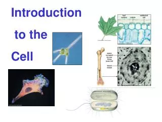
Cell Introduction
Cell Introduction. 4.1. History. Robert Hooke, in mid-1600’s, coined the phrase ‘cell’ after looking at cork b/c it looked like a monk’s room. Anton van Leeuwenhoek saw inside the cell. . cell theory:. All living things are made of cells

Cell Introduction
E N D
Presentation Transcript
History • Robert Hooke, in mid-1600’s, coined the phrase ‘cell’ after looking at cork b/c it looked like a monk’s room. • Anton van Leeuwenhoek saw inside the cell.
cell theory: • All living things are made of cells • Cells are the basic unit of structure & function of organisms • Cells come from other cells
SIZE • average plant/animal is 10 to 50 µm (micrometer = 0.0000001 or 10-6) • all nutrients enter the cell thru its surface • Greater surface area allows in more nutrients (potato demo) • As cell grows, its volume inc more rapidly than surface so, eventually it doesn’t get enough nutrients quickly enough to meet it’s needs
SHAPE • is related to function e.g. Blood cell changes shape: allows it to move thru narrow openings to isolate, engulf & destroy bacteria
Cell membrane • surrounds the cell • Regulates movement in/out of cell
Note: • Extracellular fluid – outside of cell • Cytoplasm – inside of cell Both are watery (aqueous) environments
(review) • Organelles – cell parts that perform a specific function • Nucleus – lg, near ctr, has genetic info & directs activities • Eukaryotes – (eu = true; kary = nucleus) contains membrane bound nucleus & organelles e.g. animals & plants • Prokaryotes – (pro = before; kary = nucleus) no membrane around nucleus or organelles e.g. bacterium
Membrane – regulates movement in/out of cell - Selectively permeable or semi-permeable Made of lipids & proteins
Lipid bilayer • (bi means 2) Arrangement in membrane facing outside/inside of cell • (remember - Phospholipid – head is polar/tail is nonpolar -- See fig. 4-5 on pg.74) • phospholipid – heads outward facing water environment so water can come in hydrating the cell while tails inward, keeping water inside • steroids – between phospholipids – cholesterol in animal cells • (see picture of bi-layer)
Peripheral proteins – located on the edge of bilayer membrane Integral proteins – embedded in bilayer Both used for transporting molecules thru layer
Fluid mosaic model • membrane is dynamic • acts like fluid • lipids & proteins move laterally • view short animations on membrane: http://telstar.ote.cmu.edu/Hughes/tutorial/cellmembranes/
ORGANELLES • Animal & Plant model - http://www.cellsalive.com/cells/cell_model.htm
Cytoplasm – area between membrane & nucleus • Cytosol – gelatin, aqueous fluid within cytoplasm
Mitochondria • (mitos = thread; chondrion = grain) “POWERHOUSE” • large organelle where chemical reactions that transfer energy from organic compounds to produce ATP (molecule used by cells as main energy source) • Number in cell varies – related to function of cell e.g. liver needs more b/c needs more energy • Moves within cell • Cellular respiration occurs here • Contains enzymes to catalyze chemical reactions • mtDNA (mitochondrial DNA)– grow & divide to produce new mitochondria
Smooth outer membrane – lipid bilayer – permeable to small solutes; blocks macromolecules • Cristae – inner membrane w/folds to enlarge surface area for chemical reactions to take place (cell respiration)
New Theory • – mitochondria developed because as a prokaryote it gained protection by living inside the eukaryote and in turn produced energy for the eukaryote (symbiotic relationship)
Ribosomes • site for protein synthesis – “PROTEIN FACTORY” • constructed in nucleolus • assemblage of protein & RNA(ribonucleic acid) • most numerous • not surrounded by membrane (found in prokaryotic cells too) • free/loose in cytosol or attached to endoplasmic reticulum (ER)
Ribosomes manufacture proteins based on messenger RNA (mRNA) instructions. Each ribosome reads mRNA, recruits transfer RNA (tRNA) molecules to fetch amino acids, and assembles the amino acids in the proper order.
endoplasmic reticulum(ER) • “INTRACELLULAR HIGHWAY” – path along which molecules move w/in the cell • rough ER – has ribosomes • smooth ER – no ribosomes • synthesis of steroids • regulate calcium level in muscle cells • breakdown toxic substance by liver cells
Golgi apparatus – “Post Office” – processing, packaging & secreting organelle - Modifies proteins for export
Lysosomes • “THE CUSTODIAN” • sm hydrolytic enzymes w/in membranes • Acidic enviro • digest proteins, carbohydrates, lipids, DNA, & RNA, even bacteria • Rare in plant cells
Remeber that hydrolysis breaks down the water molecule and separates a polymer into monomers, thus hydrolytic means to make eliminate water, then it becomes acidic
Cytoskeleton • network of lonG protein strands in cytosol to maintain shape • Microfilaments – small strands Actin – protein molecules linked to form polymer chain • Microtubules – lg tubes • Spindle fibers – assist in the movement of chromosomes during cell division
Cilia – many short hairlike extensions fr surface assist in movement - Line respiratory tract to trap particles in air e.g. nose hair • Flagella – few or single long extension - e.g. sperm
Nuclear matrix – gives nucleus its shape • Nuclear envelope – double membrane surrounds nucleus • Chromatin – combination of DNA & protein
Chromosomes – formed when chromatins pack together before cellular division • Nuclear pores – sm holes in nuclear envelope that allow RNA to travel to cytosol for protein synthesis • Nucleolus – ribosomes are synthesized
PLANT CELLS • Cell wall – rigid, support & protect plant Cellulose fiber embedded • Vacuoles – fluid-filled; store enzymes & metabolic wastes • Plastids – contain DNA surrounded by 2 membranes • Store starch/fats • Absorb visible light – pigments • Chloroplast – site where photosynthesis takes place • Thylakoids – membranous sacs contains chlorophyll
Difference btw animal & plant cells • Plants have & we don’t: • cell wall • Vacuoles • Plastids (where photosynthesis takes place)
Activities • cell biology - http://www.cellsalive.com/cells/cell_model.htm • cell biology worksheet needed • cell biology quiz - http://www.biologycorner.com/bio1/cellquiz.html
Organization • Tissues – organized groups of cells that carry out specific functions E.g. skin, muscle • Organ – several types of tissues that interatct to perform a specific function E.g. stomach, liver, heart • Organ system – group of organs that work together to perform a set of related tasks e.g. digestive system, respiratory system • Colonial organisms – collection of genetically identical cells that live close together E.g. volvox
Organization review from least to greatest: • Cell > tissue > organ > organ system
References • http://micro.magnet.fsu.edu/cells/animals/images/animalcell.jpg • http://www.cartage.org.lb/en/themes/sciences/zoology/AnimalPhysiology/Anatomy/AnimalCellStructure/Mitochondria/mitochondria.jpg • http://images.google.com/imgres?imgurl=http://publications.nigms.nih.gov/insidethecell/images/ch2_ribosome_proteinbig.jpg&imgrefurl=http://publications.nigms.nih.gov/insidethecell/chapter2.html&h=274&w=284&sz=35&hl=en&start=10&um=1&tbnid=pGsv2UDZt4F_6M:&tbnh=110&tbnw=114&prev=/images%3Fq%3Dribosome%26svnum%3D10%26um%3D1%26hl%3Den%26rlz%3D1T4GGIC_enUS233US233%26sa%3DN • http://images.google.com/imgres?imgurl=http://computer.act.ac.th/webproject5_2548/st/m53/division/Endoplasmic%2520reticulum.jpg&imgrefurl=http://computer.act.ac.th/webproject5_2548/st/m53/division/menu.html&h=286&w=402&sz=44&tbnid=nCYKX9rqWjAVoM:&tbnh=88&tbnw=124&prev=/images%3Fq%3Dendoplasmic%2Breticulum%26um%3D1&start=1&sa=X&oi=images&ct=image&cd=1 • http://library.thinkquest.org/C004535/endoplasmic_reticulum.html • http://library.thinkquest.org/C004535/golgi_apparatus.html • http://microscopy.fsu.edu/cells/lysosomes/images/lysosomesfigure1.jpg