Download
1 / 1
10 likes | 128 Views
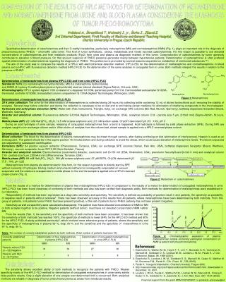
Norm. 500. 23 , 769 - HMBA. 400. Area: 11144 , 8. 13 , 727 - NMN. 300. Area: 2799 , 2. 200. 17 , 916 - MN. Area: 130 , 878. 100. 0. 10. 12 , 5. 22 , 5. 27 , 5. 20. 25. 15. 17 , 5. min. COMPARISON OF THE RESULTS OF HPLC METHODS FOR DETERMINATION OF METANEPHRINE

E N D
Norm. 500 23,769 - HMBA 400 Area: 11144,8 13,727 - NMN 300 Area: 2799,2 200 17,916 - MN Area: 130,878 100 0 10 12,5 22,5 27,5 20 25 15 17,5 min COMPARISON OF THE RESULTS OF HPLC METHODS FOR DETERMINATION OF METANEPHRINE AND NORMETANEPHRINE FROM URINE AND BLOOD PLASMA CONSIDERING THE DIAGNOSIS OF TUMOR PHEOCHROMOCYTOMA Vránková A., Škramlíková T.,Widimský J. jr., Škrha J., Jůzová Z. 3rd Internal Department, First Faculty of Medicine and General Teaching Hospital, Charles University in Prague, Czech Republic Introduction Quantitative determination of catecholamines and their O-methyl metabolites, particularly metanephrine (MN) and normetanephrine (NMN) (Fig. 1), plays an important role in the diagnosis of pheochromocytoma (PHEO) – chromaffin cells tumor. This kind of tumor synthetizes, stocks, metabolises and mostly secretes catecholamines. For this reason is possible to use elevated concentrations of catecholamines and their metabolic products (Fig.2) from urine and plasma as diagnostic markers of this tumor. Overproduction of catecholamines by tumor generally contributes to elevation of blood pressure. On that account suspicion of PHEO presence generates mainly between hypertensive patients. Determination of metanephrines is often prefered against determination of catecholamines regarding the diagnosis of PHEO. This preference is promoted by several reasons sequented on metabolism of mentioned substances1,2,3. The aim of the study was to compare the results of a HPLC with electrochemical detection method (HPLC-ED) for the determination of methanephrine and normethanephrine in bloodplasma and a HPLC with fluorescence detection method (HPLC-FLD) for the determination of the same analytes in conjugated form in urine. Both methods interpret the results in relation to the presence of PHEO. Experimental Determination of metanephrines from plasma (HPLC-ED) and from urine (HPLC-FLD) Standards: NMN ((±) normetanephrine hydrochloride), MN ((±) metanephrine hydrochloride) and HMBA (4-hydroxy-3-methoxybenzylamine-hydrochloride) used as internal standard (Sigma-Aldrich, St.Louis, USA) Chromatography:HPLC system Agilent 1100 consisted of a degasser G1379A, qarternary pump G1311A, thermostatted autosampler G1329A, thermostat controller G1330B, termostatted column compartment G 1316A (Agilent Technologies, Wilmington, USA) Determination of metanephrines from urine (HPLC-FLD) 24 h urine collection:The urine for the determination of metanephrines is collected during 24 hours into collecting bottle containing 12 mL of diluted hydrochlorid acid ( ensuring the stability of analytes). Several days before collection and during the collection is necessary to be on diet and to omit taking certain medicine for elimination of interfering compounds in the chromatogram. Hydrolysis and solid phase extraction (SPE):Water bath (Falc Instruments, Treviglio, Italy), ion exchange SPE columns (Bio-Rad, Munich, Germany), bottletop dispensers Seripettor (Brand, Wertheim, Germany) Detector and analytical column: Fluorescence detector G1316A (Agilent Technologies, Wilmington, USA), analytical column C18 - particle size 5 µm, [100x3 mm] (Sigma-Aldrich, St.Louis, USA). Mobile phase (MP): 221 mM NaH2PO4. 2H2O, 12.5 mM octane sulphonic acid, 2.5 mM sodium azide,CH3OH: deionized H2O (19 : 100), pH 3 During an acid hydrolysis of urine sample, releasing of conjugated metanephrines from a bond is happened. Acid hydrolysis is folloved by solid phase extraction (SPE). During SPE are analytes caught to ion exchange column matrix. After elution of analytes from the column bed, eluted sample is applied onto a HPLC reversed phase column. Determination of metanephrines from plasma (HPLC-ED) Blood collection:The blood for the determination of plasma metanephrines may be drawn through cannula, after fasting and being on diet (elimination of interferences). Heparin is used as an anticoagulant. The patient should be in the supine position 15 minutes before and during collection to avoid the stress, which could cause elevation of metanephrine levels. The blood corpuscles are separated by subsequent centrifugation. Extraction (SPE):24 positionvacuum extractor (Phenomenex, Torrance, USA), ion exchange SPE columns (Varian, Palo Alto, USA), bottletop dispensers Seripettor (Brand, Wertheim, Germany), vacuum evaporator Jouan RC10.22 (Saint-Herblain, France) Detector and analytical column: Electrochemical (coulometric) detector, coulometric cell E=150 mV (ESA, Chelmsford, USA), precolumn SecurityGuard [4.0x3.0 mm] and analytical column C18 - particle size 5 µm, [250x4.6 mm] (Phenomenex, Torrance, USA) Mobile phase (MP): 88 mM NaH2PO4. 2H2O, 590 µM octane sulphonic acid, 27 µM EDTA, CH3CN: deionized H2O (7.5 : 100), pH 3,25 Figure 1: Metanephrines Metanephrines from plasma are determinated in free form, for this reason is possible to directly start by SPE without necessity of hydrolysis. Eluting medium ammoniacal methanol is consequently evaporated on vacuum evaporator and the residue is resuspended in mobile phase. In the end the sample is applied onto a HPLC reversed phase column (Fig. 3). Results Figure 2: Metabolism of catecholamines From the results of a method for determination of plasma free metanephrines (HPLC-ED) in comparison to the results of a metod for determination of conjugated metanephrines in urine (HPLC-FLD) has been found closeness of conformity of both methods and also has been verified their diagnostic ability. Both methods for determination of metanephrines were established in our laboratory. Responsibility of methods has been expressed as a diagnostic sensitivity and specificity. The sensitivity is defined as probability of positive result in sick patients, the specificity as probability of negative result in patients without sickness4. There has been observed accuracy of the results from 40 patients, where metanephrines have been determined by both methods. From this group of patients, in 8 patients tumor PHEO has been present (positive), in the rest of patients tumor PHEO certainly has not been present (negative). Sensitivity as well as specificity were calculated subsequently. The patient must have elevated concentration of NMN or MN or both analytes together to be positive. Negative patients (without tumor) must have not elevated concentration NMN neither MN. From the results (Tab. 1) the sensitivity and the specificity of both methods have been calculated. It has been shown that the sensitivity of both methods has reached 100%, the specificity of methods is lower (94% for the HPLC-ED method and 80% for the HPLC-FLD). A study published previously5, which involved more extensive group of patients, shows the sensitivity and the specificity of metanephrines in plasma 99 %, resp. 89 % and the sensitivity and the specificity of metanefrines in urine 97 %, resp. 69 %. Table: The number of correctly established patients by both methods. (Total number of patients has been 40) Figure3: Chromatographic separation ofplasma metanephrines sample containing patological concentration of NMN (a patient with pheochromocytoma). References 1. Eisenhofer G., Walther M. M., Huynh T. T., Li S. T., Bornstein S. R., Vortmeyer A., Mannelli M., Goldstein D. S., Linehan W. M., Lenders J. W. M., Pacák K.: J. Clin. Endocrinol. Metab. 86, 1999 (2001). 2. Eisenhofer G., Lenders J. W. M., Goldstein D. S., Mannelli M., Csako G., Walther M. M., Brouwers F.M., Pacák K.: Clin. Chem. 51, 735 (2005). 3. Pacák K.: Inaugural dissertation. Charles University, Prague 2002. 4. http://new.euromise.org/czech/tajne/ucebnice/html/html/node5.html,downloaded in December, 10, 2007. 5. Lenders J. W. M., Pacák K., Walther M. M., Linehan W. M., Mannelli M., Friberg P., Keiser H. R., Goldstein D. S., Eisehofer G.: J. Am. Med. Asoc. 287, 1427 (2002). Conclusions The sensitivity shows excellent ability of both methods to recognize the patients with PHEO. Weaker specificity mainly of the HPLC-FLD method for detrmination of conjugated metanephrines in urine rarely admits false-positive results. Only a slight elevation of one analyte over determined limit is concerned. Both analytical methods are reliable in diagnosis of tumor pheochromocytoma as arises from introduced results. Financial support from the grant MSM 0021620807, is gratefully acknowledged.