Laser microdissection
Laser microdissection. MCB 1 st April, 2013. Purpose. To allow us to utilise material that cannot be obtained by other methods To select only the cells or tissue that we want. Overview of LMD. Excise tissue (cells) from tissue Dissection under microscope

Laser microdissection
E N D
Presentation Transcript
Laser microdissection MCB 1st April, 2013
Purpose To allow us to utilise material that cannot be obtained by other methods To select only the cells or tissue that we want
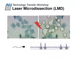
Overview of LMD • Excise tissue (cells) from tissue • Dissection under microscope • Exclude contaminating cells/materials • Many potential tissue sources • Accurate discrimination (staining, hi-mag)
Example of fine selection Honaaset al. Functional genomics of a generalist parasitic plant: Laser microdissectionof host-parasite interface reveals host-specific patterns of parasite gene expression. BMC Plant Biology 2013, 13:9
Downstream applications • QPCR, RNA-seq, microarray for gene expression analysis • Protein isolation • DNA sequencing for tissue specific sequences • More detail in the next laboratory presentation
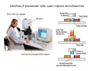
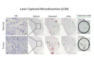
Technical overview • Attach tissue to membrane (PEN, etc.) • Dissect out desired tissue with laser • LMD is not the same as LCM. Laser Capture Microdissection Emmert-Buck et al., 1996
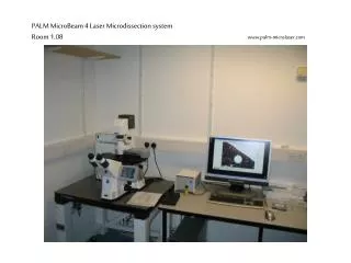
The microscope Microscope software Laser power source X-Y and objective controls Microscope
Sectioning the tissue A B C Figure 1. Laser microdissection enables profiling of gene expression at different stages of leaf development. Morphogenetic primordia (A, <750μm) were excised separately from maturing primordia (B, >750μm) and from the shoot apical meristem (remaining in C). In addition to classification of primordia by length, the area of the tissue excised is recorded. Tissues for each stage were pooled for RNA extraction using an Arcturus Paradise kit. Randomly primed reverse transcription has yielded cDNA for quantitative PCR comparison of development-associated candidate gene expression. Satisfactory yields were obtained from approximately 300 10μm tissue sections.
Harvest tissue ✓ Plant shoot tips – mature leaf and stem, young leaf and SAM Fix/preserve tissue ✓ 3:1 ethanol:acetic acid Mount tissue on membrane Leica PEN membrane slides (Heat block) Deparaffinise Ethanol/ xylene; 100% ethanol; 95% ethanol; 70% ethanol Stain material for identification E.g. eosin, haematoxylin Dissect tissue Genomics Facility LMD 6000 Downstream processing RNA, DNA, protein, carbohydrates, etc.
Protocol-specific decisions • Tissue type, quality and quantity • Fixation method- Paraformaldehyde; acetone; ethanol/ethanol:acetic acid; methacarn (methanol, chloroform and acetic acid)- Affects morphology and downstream processes • Cryogenics or paraffin embedding- Preservation of molecules or morphology • Sectioning- Section thickness; arrangement, pooling • Additional steps- RiboAmp?
References • Leica LMD home - http://www.leica-microsystems.com/products/light-microscopes/life-science-research/laser-microdissection/ • Leica LMD preparation protocol - http://www.unige.ch/medecine/bioimaging/equipment/microscopes/protocoles.pdf • Fixation methods - Kim, J.-O., Kim, H.-N., Hwang, M.-H., Shin, H.-I., Kim, S.-Y., Park, R.-W., Park, E.-Y., Kim, I.-S., Van Wijnen, A.J., Stein, J.L., Lian, J.B., Stein, G.S., Choi, J.-Y.Differential Gene Expression Analysis Using Paraffin-Embedded Tissues after Laser Microdissection(2003) Journal of Cellular Biochemistry, 90 (5), pp. 998-1006. • Plant tissue fixation - Kerk, N.M., Ceserani, T., Lorraine Tausta, S., Sussex, I.M., Nelson, T.M. Laser capture microdissection of cells from plant tissues (2003) Plant Physiology, 132 (1), pp. 27-35. • Example - Honaas, L.A., Wafula, E.K., Yang, Z., Der, J.P., Wickett, N.J., Altman, N.S., Taylor, C.G., Yoder, J.I., Timko, M.P., Westwood, J.H., dePamphilis, C.W. Functional genomics of a generalist parasitic plant: Laser microdissection of host-parasite interface reveals host-specific patterns of parasite gene expression (2013) BMC Plant Biology, p. 9. Article in Press. • Arcturus Paradise kit manual - http://www.excilone.com/client/document/manuel-paradise_344.pdf • LCM method - Laser Capture Microdissection for Analysis of Gene Expression in Formalin-Fixed Paraffin-Embedded Tissue. Ru Jiang, Rona S. Scott, and Lindsey M. Hutt-Fletcher • Overview with notes for hepatology, protein, RNA and DNA - Laser Microdissection Microscopy and its Applications in Molecular Biology. AhlamMustafa, Ragai R Mitryand Alberto Quaglia, 2010 Acknowledgements Great thanks to Vijay Nadella at the Genomics Facility (5th floor, Porter Hall). Useful discussions were had with DrFaik and with Loren Honaasfrom Penn. State.