Download
1 / 13
130 likes | 198 Views
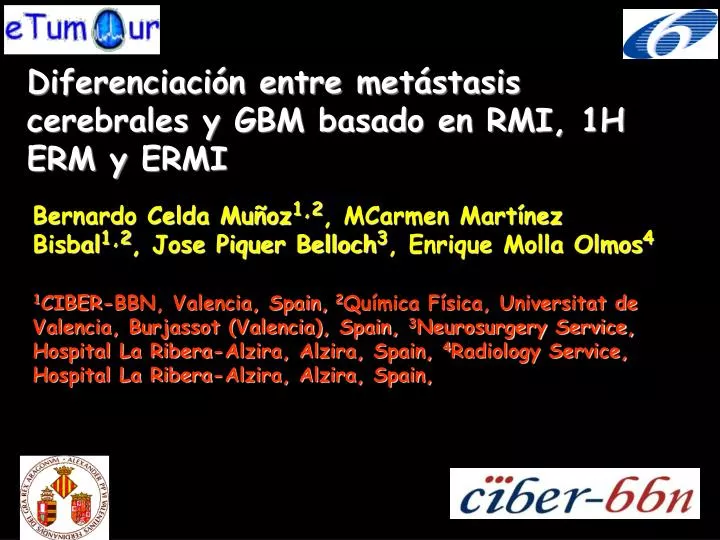
Diferenciación entre metástasis cerebrales y GBM basado en RMI, 1H ERM y ERMI. Bernardo Celda Muñoz 1,2 , MCarmen Martínez Bisbal 1,2 , Jose Piquer Belloch 3 , Enrique Molla Olmos 4.

E N D
Diferenciación entre metástasis cerebrales y GBM basado en RMI, 1H ERM y ERMI Bernardo Celda Muñoz1,2, MCarmen Martínez Bisbal1,2, Jose Piquer Belloch3, Enrique Molla Olmos4 1CIBER-BBN, Valencia, Spain, 2Química Física, Universitat de Valencia, Burjassot (Valencia), Spain, 3Neurosurgery Service, Hospital La Ribera-Alzira, Alzira, Spain, 4Radiology Service, Hospital La Ribera-Alzira, Alzira, Spain,
Objetivos El diagnóstico diferencial entre lesiones solitarias GBM y metástasis de cerebro mediante el uso combinado de espectroscopía de Resonancia Magnética (ERM), espectroscopía de Resonancia Magnética de Imagen (ERMI) y RMI.
Material y Método Datos de RMI y ERMI para 15 pacientes con lesiones solitarias (9 GBM y 6 metástasis) se adquirieron como parte de la rutina preoperativa. Imágenes RMI potenciadas en T1 (pre y post-Gd) y T2 y 2D-TSI (24x24, FOV 230x230 mm, espesor de corte 20mm, TE=272ms, TR=2000ms) se adquirieron en un Philips 1,5T. Los cocientes Co/Cr y Co/NAA se calcularon en regiones: i) sin realce en T2, ii) región peritumoral (no realce-hiperintensa en T2) y iii) realce en tumor.
RESULTS In vivo MRS Brain primary tumour vs metastases MRSI 2D TSI at 272 ms 1.5 T Brain secondary lesion metastasis (MT) (Cho/Cr)MT<(Cho/Cr)GBM Brain brimary tumour (GBM) (FP6-2002-LIFESCIHEALTH 503094) Celda et al., Adv Exp Med Biol 2006 http://www.etumour.net,
RESULTS Co In vivo MRS Brain tumour radionecrosis vs. recurrence MRSI Transverse 2D MRSI 1.5 T TE 31 ms TR 2 s Cho Tumour Recurrence (TR) (Cho/NAA)TR >(Cho/NAA)RI Sagital 2D TSI 1.5 T TE 272 ms TR 2 s Radiation injury (RI) (FP6-2002-LIFESCIHEALTH 503094) Celda et al., Adv Exp Med Biol 2006 http://www.etumour.net,
RESULTS Nosologic Image of Brain Tumour: MRI + MRSISegmentation (MRI) + Classification (MRSI) by 4 model tissue using 3 MRI slices In vivo MRS Necrosis+T Infiltration T+infil Grey M White M. LCR Laudatio, Martinez-Bisbal et al NMR Biomedicine 2007
MET – 267pmrSegmentation + Classification results obtained by 4 tissue models on the original grid and carried out on more MRI slices Dark blu = gray matter; light blue = white matter; green = csf; yellow = tumor+infiltration; brown = necrosis+tumournecrosis; gray-blue = normal+axdamage; Top right image: results obtained within the lesion detected by the segmentation algorithm Bottom left image: the white pixels represent normal tissue on MRI, but (based on CCA) tumor+infiltration on MRSI Bottom right image: the white pixels represent normal tissue on MRI, but (based on CCA) tumor+infiltration or necrosis on MRSI
Guided Therapy;Radiotherapy, Quimio Tumour tissue Infiltration A B Necrosis Normal tissue Colaboration Prof. Sabine van Huffel, Kul, Leuven T. Laudatio, MC. Martínez-Bisbal et al., NMR Biomedicine (2007)
MOLECULAR IMAGING: MRSI vs. PET B A Metastatic Cystic NAA Lac Co Cr NA
MOLECULAR IMAGING: MRSI vs. PET B A 3 1 6 4 2 7 5 C High Grade Glioma
Resultados Los valores promedio Co/Cr y Co/NAA en las regiones peritumoral y tumoral son mayores en GBM que en metástasis, lo que permite una clara separación entre ambos tipos de lesiones. La infiltración a través de imágenes nosológicas (ERMI + RMI) permite una diferenciación directa entre lesiones solitarias de GBM y metástasis.
Conclusiones El uso combinado ERMI (2D-TSI) y RMI permite la diferenciación entre GBM y mestástasis en lesiones solitarias mediante: a) cocientes Co/Cr y Co/NAA en las diferentes regiones de la lesión b) la naturaleza infiltrativa de GBM detectada por imágenes nosológicas