Download
1 / 24
250 likes | 996 Views
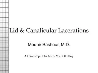
Lid & Canalicular Lacerations. Mounir Bashour, M.D. A Case Report In A Six Year Old Boy. Introduction. A short presentation to stimulate a discussion on a practical approach to complex lid/canalicular lacerations.

E N D
Lid & Canalicular Lacerations Mounir Bashour, M.D. A Case Report In A Six Year Old Boy
Introduction • A short presentation to stimulate a discussion on a practical approach to complex lid/canalicular lacerations. • By Mounir Bashour, PGY-3, Ophthalmology, George Washington University, graduate of McGill Medical School.
Case Presentation/HPI • 6 yo bm presents with complex lid laceration OS. • Secondary to falling from upper bunk bed while playing around 2 AM 7/20/95. • Hx of Prematurity (28 weeks) was in NICU for 3 months, no Hx of ROP. • Currently good health, no meds, allergies • Single parent (father) family.
Examination • >4 cm full thickness medial oblique upper lid laceration OS extending into medial canthus. • PERRLA, no RAPD. • Va 20/30 OU by Snellen. • Rotations full, ortho. • No corneal abrasion, Seidel negative. • Dilated exam reveals picture consistent with resolved early ROP.
Photo of Upper Lid Laceration • Photo with similar laceration as found in our patient.
Diagnosis • Suspicion • Common etiologies • Epidemiology
Necessity of Repair • Controversy • Jones study • Moore and Linberg study
Timing of Repair • Immediate vs late
Discussion I • The aim of lid repair • Workup
Discussion II • Blunt injuries
Discussion III • Lacerations involving the canthal angles
Intraoperative Complications • Inabilty to Locate the Medial End of the Canaliculus • Difficulty Retrieving Probe from Nose • Problems Suturing the Canalicular Walls • Difficulty Repairing Medial Canthal Ligament Injury
Proximal Canaliculus • The characteristic appearance of the proximal canaliculus
Normal Anatomy of the Lacrimal System • Essential knowledge
Intubation • Gavaris Modification of the Quickert-Dryden procedure
Anastamosis of the Canaliculus • Problems with suturing
Medial Canthal Ligament Injury • Correct Placement of MC Fixation Suture • (A) Posterior reflection of MCT behind the lacrimal sac • (B,C) Correct fixation point
Intubated Nasolacrimal System • Double-knotted Silastic Tubing
Complications With Silicone Tubes • Tube displacement • Punctal/canalicular erosion/slitting • Conjunctival/corneal irritation • Granuloma formation • Epistaxis
Displaced Tubing • Most common complication
Securing the Tubing • One method of several
Erosion • Six knots with 4-0 nylon woven into knots • Secured to lateral vestibule of nose
Granuloma • Granuloma formation from silicone tubing • Displaced silicone tubing after patient had caught tubing with finger and pulled loop onto cheek
Rarer Complications • Dacryocystitis • Epiphora • Ectropion • Loss of tubing • Difficulty removing tubing