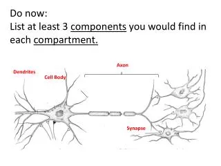
Differences Between axon and dendrites
Differences Between axon and dendrites. Axon:(efferent) 1-single process 2-thin&long 3-uniform diameter along its length 4-branches&also give collateral 5-contains neurofibrils but no nissl granules 6-Two types of axonal transport exist Antigrade&retrograde. The Dendrites(Afferent).

Differences Between axon and dendrites
E N D
Presentation Transcript
Differences Between axon and dendrites • Axon:(efferent) • 1-single process • 2-thin&long • 3-uniform diameter along its length • 4-branches&also give collateral • 5-contains neurofibrils but no nissl granules • 6-Two types of axonal transport exist Antigrade&retrograde
The Dendrites(Afferent) • 1-usually multiple process • 2-usually short&thick • 3-their thickness decreases gradually towards its end • 4-have many fine side projection called spines • 5-Contain Nissl granules&neurofibrils • 6-receive impulses from other neurones
The Axon • Formed of cytoplasm known as axoplasm containing • mitochondria,neurofibrils&neurotubule • surround by membrane(axolemma)
Classification of neurons • -Golgi type 1neuron:They have long axons as the neurons of cerebral,cortex • -Axons from the tracts of the brain&spinal cord • -Golgi type2 neuron:have short axons as neurons of retina
Classified axon according to covering sheates • -The axon covered with myelin sheath or neurolemma • -1-Naked nerve axons without amyelin sheath without aneurolemma as found in grey matter • -2-covered with myelin sheats but without neurolemma found in whait matter
3-nerve fibres covered with myelin sheaths&covered aso with neurolemma found in outside the spinal cord • 4-covered with neurolemma but are not covered with myelin sheaths found in post ganglionic sympathetic nerve axon
The Myelin Sheaths • -Fatty tubular covering around the axon • -formed by neurolemma cells surround the peripheral nerves • -The the brain formed by neurglia cells(oligodendroglia) • -composed of cholesterol,fatty acids&phospholipids
MYELIN MESAXON MYELIN SHEATH Schwann cell AXON cytoplasm major dense interperiod line
SHEATH OF SCHWANN node of Ranvier internodal region clefts of collateral Schmidt-Lanterman Schwann sheath axon
-Interrupted at intervals by nodes of renvier&lantrermanns clefts • -Lantermanns clefts:discontinuities in the myelin sheath, • facilitate the passage of the nutrition from schwann cells to the myelin sheath
Function of myelin sheath: • - Protection of the axons • -accelerate,conduction of nerve impulse • -isolates nerve impulses
Schwann cells(neurolemmal) • -formed of achain of schwan cells around M.Sheath • -Each cell corresponds to an inter nodal segment&its comes in contact with axon at the nodes of ranvier
-Schwan cells in order to form the myelin sheath around the axon • -encircles the axon&rotates several tymes around it&forming series of rings
Function of neurolemmal cells • 1-isolate nerve impulses • 2-help in regeneration of neurons • 3-form the myelin sheath around axons
The Synapse • The point of contact between the processes of the neurons • -there is no cytoplasmic continuity between neurons • -Synapses may be Excitatory Or Inhibitory • -Chemical synapse transmit impulses through neurotransmitters • -Electrical synapse transmit impulse directly
Types of Synapses • a-Axo-dendritic synapse:contact between the axon of one neuron&dendrites of another neuron • b-Axo-somatic synapse&cell body of another neuron • C-Axo-axonic synapse:contact between the axons of 2 neurons • d-Dendro-dendritic:contact between dendrites of 2 neurones
Function: • a-Excitaory or inhibitory • b-transmit impulses through neurotransmitters • c-transmit impulse directly • d-contact between the processes of the neurons
The structure of aperipheral nerve trunk • Formed of collections of axons arranged in bundles • -nerve trunk is surrounded by C.T. fascia called • -Epineurium • -Perineurium • -Endoneurium(Henls Sheath)
PERIPHERAL NERVE TRUNK Nerve Trunk Schwann Cell nuclei Endoneurium (collagen fibers, fibroblasts) Epineurium (epitheloid & myoid cells) Perineurium (dense connective tissue) fasicle
Nerve trunk stained with osmic acid,the myelin sheaths will appear as black circles(osmic acid stains only the myelin sheath)
PERIPHERAL NERVES (Osmic Acid) Epineurium Fasicle Fasicle Perineurium Fasicle Myelin Sheath Axon
Nerve Ganglia • Formed of nerve cells&nerve fibres surrounded by C.T. • -there are two types of ganglia • 1-Spinal ganglia • 2-Autonomic ganglia • a-sympathetic • b-Parasympathetic
1-spinal ganglia • Present beside the spinal cord • -They act as relay for sensations: • -Formed of pseudo-unipolar nerve cells • -each nerve cell is surrounded by many supportive satellite cells • -the ganglion is covered by thick capsule
2-Sympathetic ganglia • present as isolated ganglia in the different regions of the body • -formed of multipolar stellate-shaped nerve cells
Neuroglia • -There is no C.T. in CNS • -they form the supporting tissue between the neurons of CNS • Stained with silver or gold choloride
Classification of neuroglia • 1-neuroglia proper have 3 types • a-macroglia or astrocytes(protoplasmic and fibrous) • b-microglia(mesodermal in origin) • c-oligodendroglia(with few dendrites)
2-other types of supporting neuroglia-like cells • Present in nervous tissue • a-Epedymal cells • they are derved from the spongioblast cells • -they form the (CSF) • b-Satellite cells :they are the small cells surround the nerve cells,have nutritive functions to nerve cells
c-schwan cells: • present around the axons of the peripheral nerves form myelin sheath..help in regeneration • d-spongiblast cells: primitive embryonic cells which can differentiate into neuroglia cells • e-Tancyte cell:which surround the neurons of hypothalamus
The Neuroglia Proper • 1-Macroglia or Astrocytes(star Shaped) • There are 2 types of astrocytes • A-protoplasmic astrocytes • B-Fibrous astrocytes
A-Protoplasmic astrocytes • -arising from spongioblast • -present in the grey matter of CNS • -they are branched cells • -ctoplasm rich in granules known as gliosomes • -they can divide
B-Fibrous astrocytes • -similar to protoplasmic astrocytes • -present in the white matter of CNS • -cytoplasm non granular
2-Microglia or Mesoglia • They are mesodermal in origin • -they are present in grey&white matter • -they are small spindle-shaped cells • -they have nissls granules • They are phagocytic cells during inflammation&during degeneration®eneration of neurons • -they play defensive role during AIDS disease
3-oligodendroglia(few dendroglia) • -present in the grey &white matter • -small branched cells with large deeply stained nuclei • They can form the myelin sheats • They present around B.V.&nerve cells
GREY AND WHITE MATTER Grey Matter WHITE AND GREY MATTER nerve cell bodies nerve white matter glial fibers nuclei White Matter dura axons grey matter mater (fibers) arachnoid membrane motor glial neurons pia nuclei mater
NEUROGLIA MICROGLIA (mononuclear phagocytic system) OLIGODENDROGLIA (forms Scwann sheath of CNS)
Functions of neuroglia • 1-support the neurons • 2-form the myeline sheats • 3-separating the neurons • 4-nutritive functions for neuroned • 5-defend against inflammation of neurons • 6-regeneration of neurons • 7-secrete the CSF • 8-the secrete Enkephlin to influence neuronal activity
Degeneration®eneration of neurons • Cutting of an axon of neurons(knife,trauma or infection • Result in: • 1-Retrograde degeneration(nerve cell) • 2-Traumatic degeneration(site of injury) • 3-Wallerian degeneration(peripheral part of the axon)
Retrograde Degeneration • Changes in shape,size nucleus,cell organoids&cell inclusion of the nerve cell • The nerve cells,chnges:swells,loses dendrites ,small in size,cytoplasm pale,nucleus pyknotic nuclear chromatolysis,disappearance of nissl granules
-nissl granules: veryy sensitive to any toxin • Nissl granules can be observed by methylene blue or by hematoxylin stain
Neurofibrils(golgi appartus) degenerate gradually&finally disappear(stained with silver stain,it will lead to complete death of the nerve cells
Wallerian Degeneration • Changes occure in the axon,myelin sheath&neurolemma after partial cutting of the axon • 1-chnges in the axon; neurofibril are swollen at interrupted areas giving the axon • The axon begins to break into small pieces of different length which can stained in silver
2-changes in the myelin sheaths • -segmention to form fermention chambers • -the normal shape of M.Sheath in change into droplets of neural fat7oliac acids can stained in osmic acid • 3-chnges in the neurolemma • -increase in zise&nuclei divide by mitosis the cells form atubular cord around the myelin sheath helping in regeneration of the cut neurons
N.B.:traumatic degeneration • rapid process of W.degeneration
Regeneration of neurons • Depends up on the degree of nerve injuries&up on these conditions • -grade of cutting(partial ,complete0 • -depends upon the distance between the two cut ends • -wound infected or not • -neurolemma is intact or not
-If there is no infection in partial cut&neurolemma intact&the neurofibrils of the central stump will grow,these fibrils are known as aton sprouts • -regeneration in human nerve occurs slowly
Transneural degeneration • The neuron are functionally connected by synapse,if there is cutting,there is no stimuli from the other neurones
NERVE CELL BODY myelinated axon axo-denritic synapse unmyelinated axons axo-somatic synapse axon hillock neurotubules neurofilaments axon vesicles (neurotransmitters) mesaxon axon