
Antibiotic Sensitivity Testing
E N D
Presentation Transcript
Antibiotic Sensitivity Testing Dr.T.V.Rao MD
Uses of Antibiotic Sensitivity Testing • Antibiotic sensitivity test: A laboratory test which determines how effective antibiotic therapy is against a bacterial infections. • Antibiotic sensitivity testing will control the use of Antibiotics in clinical practice • Testing will assist the clinicians in the choice of drugs for the treatment of infections. • Dr.T.V.Rao MD
Components of Antibiotic Sensitivity Testing • 1.The identification of relevant pathogens in exudates and body fluids collected from patients • 2. Sensitivity tests done to determine the degree of sensitivity or resistance of pathogens isolated from patient to an appropriate range of antimicrobial drugs • 3. Assay of the concentration of an administered drug in the blood or body fluid of patient required to control the schedule of dosage. Dr.T.V.Rao MD
Antibiotic Sensitivity Testing Is Essential of selection of Antibiotics
Isolation and Identification of Bacteria precedes the selection of Antibiotic Testing Methods
Uses of Antibiotic Sensitivity Testing • Helps to guide the Physician in choosing Antibiotics • The accumulated results on different pathogens their sensitivity will guide the physician in choosing empirical treatment in serious patients before the individual’s laboratory results are analyzed in the Microbiology laboratory. • Reveals the changing trends in the local isolates. • Helps the local pattern of antibiotic prescribing. • Dr.T.V.Rao MD
Why Need continues for testing for Antibiotic Sensitivity • Bacteria have the ability to develop resistance following repeated or subclinical (insufficient) doses, so more advanced antibiotics and synthetic antimicrobials are continually required to overcome them. • Dr.T.V.Rao MD
Testing for Antibiotic sensitivity • The method includes several steps including obtaining a bacterial sample; identifying the type of bacteria in the bacterial sample; selecting a set of antibiotics based on the identity of the bacteria in the bacterial sample; obtaining a control sample from the bacterial sample; Dr.T.V.Rao MD
Testing Antibiotic Susceptibility • Antibiotic sensitivity test: A laboratory test which determines how effective antibiotic therapy is against a bacterial infections. • Antibiotic sensitivity test: the in vitro testing of bacterial cultures with antibiotics to determine susceptibility of bacteria to antibiotic therapy. Bauer-Kirby test. Dr.T.V.Rao MD
How results to be Reported after Sensitivity Testing • One should follow guidelines in reporting the results and make matters simple with clear words as Organism A isolated and sensitive ( or resistant ) to Antibiotic B Such a report is relevant to present clinical condition “ that the minimum inhibitory concentration( MIC ) of the antibiotic for it has been measured in some way and that, if the organism is reported as sensitive the MIC is less than a half or quarter of the concentration of antibiotics likely to be found in the infected tissues of a patient given the usual schedule of doses i.e. that the infection is treatable”
What is Resistance in Antibiotic Sensitivity Testing • Resistance implies that the infection is not treatable with the tested Antibiotic because its MIC exceeds achievable safe tissue or urine levels. • Intermediate sensitivity means they show aunimodal distribution, but they can often be reclassified as sensitive or resistant if retested. • Dr.T.V.Rao MD
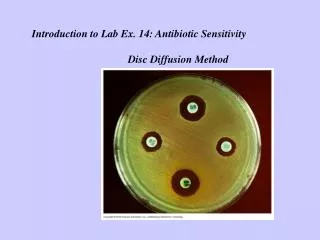
Kirby-Bauer methods A commonly used method in basic laboratories • Kirby-Bauer antibiotic testing (KB testing or disk diffusion antibiotic sensitivity testing) is a test which uses antibiotic-impregnated wafers to test whether particular bacteria are susceptible to specific antibiotics • Dr.T.V.Rao MD
How to perform Kirby- Bauer testing • The basics are easy: The bacterium is swabbed on the agar and the antibiotic discs are placed on top. The antibiotic diffuses from the disc into the agar in decreasing amounts the further it is away from the disc. If the organism is killed or inhibited by the concentration of the antibiotic, there will be NO growth in the immediate area around the disc: This is called the zone of inhibition. • Dr.T.V.Rao MD
Streaking the Inoculum • Kirby-Bauer (also Bauer-Kirby) disk diffusion antibiotic susceptibility testing applies a defined inoculum (compared to McFarland 0.5 OD standard streaked as a lawn onto a large Mueller-Hinton agar or Blood agar plate (in 3 directions to ensure confluence). • Dr.T.V.Rao MD
Making proper inoculum • Swab a Mueller-Hinton plate with each of the bacteria. Dip a sterile swab into the broth and express any excess moisture by pressing the swab against the side of the tube. Dr.T.V.Rao MD
Bacteria are inoculated as lawn culture • Method of inoculation- Good results are obtained by placing a standard loopful of inoculum suspension on the plate and then spreading it with a dry sterile swab.
Disk Diffusion Method • After completely swabbing the plate, turn it 90 degrees and repeat the swabbing process. (It is not necessary to re-moisten the swab.) Run the swab around the circumference of the plate before discarding it in the discard bag. • Dr.T.V.Rao MD
Placing the Antibiotic disks • Then, using a dispenser such as the one pictured, antibiotic-impregnated disks are placed onto the agar surface. As the bacteria on the lawn grow, they are inhibited to varying degrees by the antibiotic diffusing from the disk • Dr.T.V.Rao MD
Zone sizes differ on sensitivity pattern • It has been determined that zones of inhibition of a certain diameter (varies for antibiotic and to a lesser extent, bacterial species) correlate with sensitivity or resistance to the antibiotic tested
Look at the Charts for establishing the zones of Sensitivity • The zone sizes are looked up on a standardized chart to give a result of sensitivie, resistant, or intermediate. Many charts have a corresponding column that also gives the MIC (minimal inhibitory concentration) for that drug.
Interpretation • Place the metric ruler across the zone of inhibition, at the widest diameter, and measure from one edge of the zone to the other edge. HOLDING THE PLATE UP TO THE LIGHT MIGHT HELP. • The disc diameter will actually be part of that number. If there is NO zone at all, report it as 0---even though the disc itself is around 7 mm. • Zone diameter is reported in millimetres, looked up on the chart, and result reported as S (sensitive), R (resistant), or I (intermediate).
The area of Inhibition is measured with a Scale • Record the results for everyone on your table in the table below.
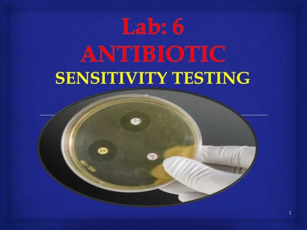
The disk diffusion methods are commonly used for routine testing
The zone of inhibition guides the right choice of Antibiotic
How results to be Reported after Sensitivity Testing • One should follow guidelines in reporting the results and make matters simple with clear words as Organism A isolated and sensitive ( or resistant ) to Antibiotic B Such a report is relevant to present clinical condition “ that the minimum inhibitory concentration( MIC ) of the antibiotic for it has been measured in some way and that, if the organism is reported as sensitive the MIC is less than a half or quarter of the concentration of antibiotics likely to be found in the infected tissues of a patient given the usual schedule of doses i.e. that the infection is treatable”
Stokes’ Method • In original Stokes’ method the inoculum of the control strain is evenly spread over the upper and lower thirds of a plate and that of the test strain over the central third;uninoculated gaps 2 – 3 mm wide are left to test from the control areas. • Dr.T.V.Rao MD
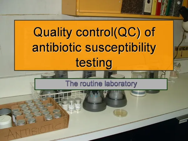
Control strainsin Stokes’ Method • The Microbiology laboratories should always practice to test the quality control of their work with the use of standard strains in Stokes method with following standard strains and results are compared 1 Escherichia coli NCTC 10418 2 Pseudomonas aeruginosa NCTC 10662 3 Staphylococcus aureus NCTC 6571
Stokes Method for Antibiotic Sensitivity testing • In the Stokes controlled sensitivity test, a control organism is inoculated on part of a plate and the test organism is plated on the remainder. Disks are placed at the interface and the zones of inhibition are compared. The use of a sensitive control shows that the antibiotic is active, so that if the test organism grows up to the disk it may safely be assumed that the test organism is resistant to that drug. • Dr.T.V.Rao MD
The strips with multiple Antibiotics can be tested in one go
Other methods of Antibiotic susceptibility testing • Other methods to test antimicrobial susceptibility include the Stokes method, E-test (also based on antibiotic diffusion). Agar and Broth dilution methods for Minimum Inhibitory Concentration determination.
Testing Minimum Inhibitory Concentration • In alternative measure of susceptibility is to determine the Minimum Inhibitory Concentration (MIC) and the Minimum Bactericidal Concentration (MBC) of a drug. A series of broths are mixed with serially diluted antibiotic solutions and a standard inoculum is applied. After incubation, the MIC is the first broth in which growth of the organism has been • Dr.T.V.Rao MD
What is Minimum Inhibitory concentration • Minimum inhibitory concentration (MIC), in microbiology, is the lowest concentration of an antimicrobial that will inhibit the visible growth of a micro organism after overnight incubation. Minimum inhibitory concentrations are important in diagnostic laboratories to confirm resistance of micro organisms to an antimicrobial agent and also to monitor the activity of new antimicrobial agents.
The Antibiotics are diluted to various dilution to test the minimum inhibitory concentration
What is E Test • Etest is an antimicrobial gradient technique in which 15 reference MIC dilutions of an antibiotic have been repackaged with innovative dry chemistry technology onto a plastic strip. The predefined gradient provides precise and accurate assessment of antimicrobial activity against both fastidious and non-fastidious microorganisms. • Dr.T.V.Rao MD
The strips are impregnated with various concentration of Antibiotics
Multiple drug resistant organisms Multiple drug resistant organisms are resistant to treatment with several, often unrelated, antimicrobial agents as described above in Shigella. Some of the most important types of multiple drug resistant organisms that have been encountered. Dr.T.V.Rao MD
Examples of Bacteria of Clinical Importance • MRSA - methicillin/oxacillin-resistant Staphylococcus aureusVRE - vancomycin-resistant enterococciESBLs - extended-spectrum beta-lactamases (which are resistant to cephalosporins and monobactams)PRSP - penicillin-resistant Streptococcus pneumoniae • Dr.T.V.Rao MD
Detection of MRSA made simple with Disk Diffusion Methods • The MRSA among the Staphylococcus aureus needs detection in special condtions either by carrying out the test on medium containing 5 % Nacl with Methicillin disk with 5 or 10 micrograms • Recently the detection was made simple with testing on Oxacillin or Cefotixin disks
Newer Methods in MRSA detection • Recently developed chromogenic media combine primary growth and selectivity with differentiation from coagulase negative staphylococci. These media show improved specificity when compared with traditional media. Sensitivity is also improved but requires 48hrs incubation to achieve >85%.
Chromagar for detection of MRSA • BBL™ CHROMagar™ MRSA is a selective and differential medium for the qualitative direct detection of nasal colonization by Methicillin resistant Staphylococcus aureus (MRSA) to aid in the prevention and control of MRSA infections in healthcare settings. The test is performed on anterior nares swab specimens from patients and healthcare workers to screen for MRSA colonization. BBL CHROMagar MRSA is not intended to diagnose MRSA infection nor to guide or monitor treatment for infections
Methods to detect ESBL producers • Various combination of Antibiotic disks are used for the detection of ESBL producers in Gram negative bacilli.
Antibiotic disks helps in detection of ESBL producers • Various Antibiotic disks kept at specified place helps in detection of ESBL producers