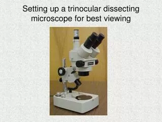
Comprehensive Microscope Guide: from Dissecting to Phase Contrast
This detailed guide covers the usage, maintenance, and activities related to dissecting and phase contrast microscopes. Learn about parts, cleaning techniques, activity preparation, and understanding phase contrast microscopy. Explore the differences between light microscopes and dissecting microscopes, oil immersion usage, total magnification calculation, and preparing slides of various specimens like animal and plant cells.

Comprehensive Microscope Guide: from Dissecting to Phase Contrast
E N D
Presentation Transcript
Storing The Microscope • Return the lowest power objective in place • Wrap the cord around the base • Return dustcover
Cleaning the Microscope • Use lens paper on all glass parts of the microscope.
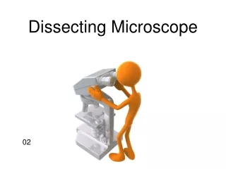
Dissecting Microscope & Parts Free standing illuminator
Pond Water Sample Paramecium Euglenia Clamydomonas Volvox Rotifer Daphnia Spirogyra
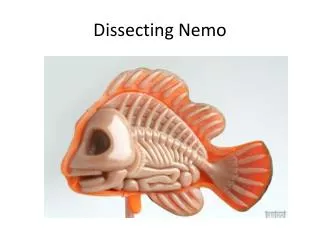
Activity • Prepare wet mount for dissecting scope • Pond water • Look at specimen under high power and draw what you see. • Use proper clean-up technique
Phase Contrast Microscope Muscle tissue Downy mildew of grape
Understanding Phase Contrast Microscopy • Imperfections in glass • Peaks and troughs don’t line up • These waves are in different phases • PCM used to transform phase differences into intensity differences, thus increasing contrast glass waves light light
Understanding Phase Contrast Microscopy Constructive interference (Light phase) Destructive interference (Dark phase)
Comparison of Light Microscopy Dark Field Bright Field Phase contrast
ocular Revolving nose piece Specimen holder objective stage Aperture iris diaphragm ring condenser Light intensity lever Y-axis knob X-axis knob Light intensity preset button Fine adjustment knob Course adjustment knob
Cleaning the Microscope • Use lens paper on all glass parts of the microscope. • Clean oil immersion lens with chemicals provided by your instructor
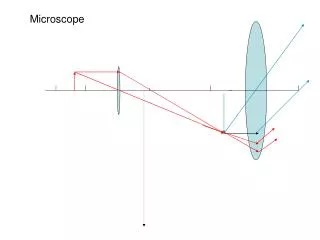
Using the microscope • Always observe using the LOWEST POWER objective first. • Focus using the COARSE ADJUSTMENT KNOB to bring the object into focus. Bring the object into sharp focus by using the fine adjustment knob. • Focus, and then move to a higher power objective, if needed. • Use only the FINE ADJUSTMENT KNOB when using the HIGHEST (longest) POWER OBJECTIVE. • Keep both eyes open to reduce eyestrain. • Determine total magnification of the object by multiplying the power of the ocular (10x) the power by the power of the objective.
Preparing a slide • Using a pipet or dropper, add a drop of water or another solvent to a clean microscope slide. Then, place the specimen in the water. • Place the edge of a coverslip on the slide so that it touches the edge of the water. • Slowly lower the coverslip to prevent the formation of air bubbles.
Oil Immersion Lens 100x 40x
ocular Nose piece objective Specimen holder stage Illumination intensity Condenser X-axis knob Iris diaphragm (aperture diaphragm ring) Fine adjustment base y-axis knob Course adjustment
Activity • Prepare wet mount • Cheek cell • Elodea cell • Onion cell • Bacterial cells in yogurt • Look at specimen under high power and draw what you see. • Use proper clean-up technique
Microscope Quiz • Which objective uses oil? • If your ocular is 10x and your objective is 40x what is the total magnification of your image? • Why do you need a cover slip and how do you avoid an air bubble? • What is the difference between a light microscope and a dissecting microscope? 1.
Aligning rings • When looking through the ocular you will see 2 rings • The may or may not be concentric. • By turning the centering adjustment screws o the condenser, you align the rings so they become concentric
Brightfield –absorption • Light is transmitted through the sample. Only useful for specimens that can be contrasted via dyes. Very little contrast in unstained specimens. • Darkfield -scattering • The illuminating rays of light are directed from the side so that only scattered light enters the microscope lenses, consequently the cell appears as an illuminated object against the view. • Phase Contrast- phase interference • Incident light [Io] is out of phase with transmitted light [I] and when the phases of the light are synchronized by an interference lens, a new image with greater contrast is seen
Dark-Field Microscopy • Modified condenser contains disc in center • Only light refracted by specimen can enter objective. • Objects surrounded by halo. • Advantage can see smaller objects like spirochetes, internal structures highlighted. • Disadvantage objects look bigger than they actually are.
Phase Contrast Microscopy • Light passes through annular diaphram. • Causes light waves to become “in phase” or synchronized. • Advantage can highlight internal cell structures and details. • Disadvantage - none.