
Exercise Physiology: Ventilatory and Cardiovascular Systems
E N D
Presentation Transcript
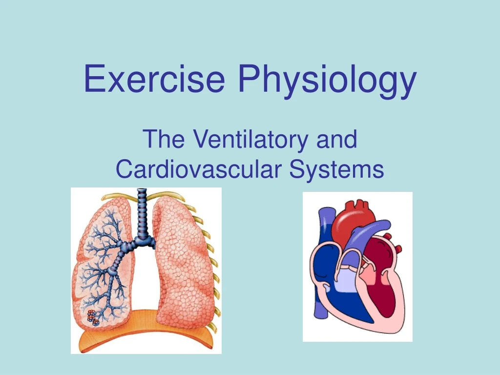
Exercise Physiology The Ventilatory and Cardiovascular Systems
I.Structure of the Ventilatory System A. Conducting Airways: *offers a low resistance pathway for air flow *warms and moistens air *mucus and ciliated cells filter air
II. Pulmonary Ventilation: the exchange of air between the atmosphere and lungs. A. Mechanics of Breathing: 1. Inhalation: *diaphragm contracts and lowers *chest cavity expands increasing volume and decreasing internal air pressure
2. Exhalation: *Diaphragm relaxes and moves up. *chest cavity volume decreases and internal air pressure increases. *during exercise the intercostal and abdominal muscles act on the ribs to produce greater exhalation
III. Total Lung Capacity: (TLC) maximum volume of lungs after maximum inhalation (vital capacity + residual vol.). • Tidal Vol.: (TV) volume inspired or expired per breath. • Inspiratory Reserve Vol.: max. inspiration at the end of tidal inspiration (Vital cap.= TV+IRV+ERV)
C. Expiratory Reserve Vol.: max. expiration at the end of tidal expiration. D. Residual Vol.: (RV) volume in lungs after maximum expiration.
IV. CO2 transport in the blood: • Dissolved directly in plasma • Bound to hemoglobin (carbamino compunds) • As bicarbonate HCO3
D. What’s the role of CO2 in the control of pulmonary ventilation during exercise? *dissociation of carbonic acid increases H+ in blood lowering pH. *the medulla senses the low pH and sends signals to the diaphragm and intercostal muscles.
V. Oxygen Transport in the Blood: • Hemoglobin: (Hb) iron containing pigment that binds with oxygen to form oxyhemoglobin. Hb + 4 O2 Hb4O8
VI. Gas Exchange in the lungs: • Alveoli: thin membrane sacs at the end of the bronchioles. *serve as the site of gas exchange by diffusion.
VII. Blood: transport vehicle for nutrients, hormones, waste products and electrolytes. 1. Blood Composition: A. Cellular: i. erythrocytes: (RBC’s) Contain hemoglobin that binds to oxygen for transport to tissues.
ii. Leukocytes: (WBC’s) defend the body against disease. *produce antibodies *destroy bacteria and viruses *produce marker proteins
iii. Platelets: (thrombocytes) play a role in the clotting of blood.
B. Liquid Component: i. Plasma: 60% total volume of blood. 90% water and 10% solutes • Metabolites and wastes (gases, hormones, vitamins) • Salts (ions) • Plasma proteins
A. Heart Rate: is regulated by both intrinsic and extrinsic factors. • Intrinsic regulation: a. sinoatrial (S-A) node: a mass of specialized cardiac muscle located on the exterior wall of the right atrium. Initiates the electrical impulse.
b. Atrioventricular (A-V) node: receives impulse from the S-A node and delays it about .10 sec. for atrial contraction. c. A-V Bundle of His: speeds the impulse over the ventricles to the Purkinje system causing simultaneous contraction of the ventricles.
ii. Extrinsic Regulation: the autonomic nervous system can override the myocardial rhythm. • Sympathetic Influence: epinephrine is released when stimulated causing heart rate to increase. • Parasympathetic Inf: releases acetylcholine to slow heart rate.
B. Circulation of Blood: i. Pulmonary Circulation: deoxygenated blood is pumped from the right side of the heart through the pulmonary arteries to the lungs. Oxygenated blood is returned by the pulmonary veins.
ii. Systemic Circulation: oxygen rich blood is pumped from the left side of the heart through the aorta to the rest of the body.
iii. Cardiac Output: the volume of blood pumped by the heart in one minute. Equal to stroke vol. x heart rate. • Stroke Vol.: the volume of blood pumped by one ventricle with each beat. Approx. 70 ml. Stroke vol.=EDV-ESV
iv. Cardiovascular Drift: an increase in heart rate during steady exercise due to a reduction in stroke volume. Caused by: *exercising in heat *rise in core temp. *decrease in plasma vol.
C. Blood Pressure: the pressure exerted on the walls of the arterial system. • Systole: highest pressure generated by the left ventricular contraction. Approx. 120 mm Hg at rest. • Diastole: the pressure generated when the heart relaxes. Approx 70-80 mm Hg.
iii. Blood Pressure Response to Exercise: • Dynamic Exercise: systolic pressure increases with intensity with relatively little change in diastolic pressure. Ex. Walking, jogging, swimming, cycling.
b. Static Exercise: heavy resistance training increases blood pressure due to muscular contractions compressing peripheral arteries. Ex. Weightlifting, isometrics
Rest (cardiac output 5,000 ml) *liver = 1350 ml *kidneys = 1100 ml *muscle = 1000 ml *brain = 700 ml *skin = 300 ml *heart = 200 ml Exercise (cardiac output 25,000 ml) *liver = 500 ml *kidneys = 250 ml *muscle = 21,000 ml *brain = 900 ml *skin = 600 ml *heart = 1000 ml iv. Distribution of Blood
v. Cardiovascular Adaptations to Exercise: • Lower resting heart rate. • Increased left ventricular volume. • Increased stroke vol. and cardiac output. • Capillarization: increase in capillary surface area in muscles. • Greater arteriovenous oxygen diff. (a-vO2)
D. Maximal Oxygen Consumption: (VO2) refers to the maximum amt. of O2 that an individual can utilize during maximal training. *measured as ml of O2 used in one minute per Kg of body weight. (ml Kg-1 min-1)