
Eclipse® Soft Tissue Anchor - Enovis Eclipse®
Eclipseu00ae Soft Tissue Anchor is used in tenodesis or tendon transfer procedures to reliably fixate soft tissue inside a bone tunnel.<br><br>Soft tissue that can be inserted and tensioned using a blind hole or pull-through technique. One-sided, the compressed profile allows the Sheath to be placed easily alongside soft tissue inside the tunnel and is loaded on a disposable Deployment Gun for fast, easy insertion and handling. Contact us at 800.456.8696 to know more or visit us at https://www.djoglobal.com/.

Eclipse® Soft Tissue Anchor - Enovis Eclipse®
E N D
Presentation Transcript
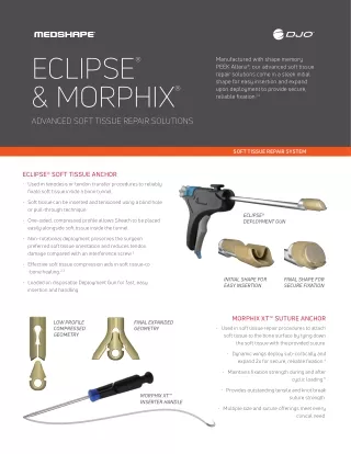
ECLIPSE & MORPHIX ADVANCED SOFT TISSUE REPAIR SOLUTIONS ® Manufactured with shape memory PEEK Altera®, our advanced soft tissue repair solutions come in a sleek initial shape for easy insertion and expand upon deployment to provide secure, reliable fixation.1,4 ® SOFT TISSUE REPAIR SYSTEM ECLIPSE® SOFT TISSUE ANCHOR • Used in tenodesis or tendon transfer procedures to reliably fixate soft tissue inside a bone tunnel. • Soft tissue can be inserted and tensioned using a blind hole or pull-through technique. ECLIPSE® DEPLOYMENT GUN • One-sided, compressed profile allows Sheath to be placed easily alongside soft tissue inside the tunnel. • Non-rotational deployment preserves the surgeon preferred soft tissue orientation and reduces tendon damage compared with an interference screw.2 • Effective soft tissue compression aids in soft tissue-to -bone healing.2,3 INITIAL SHAPE FOR EASY INSERTION FINAL SHAPE FOR SECURE FIXATION • Loaded on disposable Deployment Gun for fast, easy insertion and handling. MORPHIX XT™ SUTURE ANCHOR LOW PROFILE COMPRESSED GEOMETRY FINAL EXPANDED GEOMETRY • Used in soft tissue repair procedures to attach soft tissue to the bone surface by tying down the soft tissue with the provided suture. • Dynamic wings deploy sub-cortically and expand 2x for secure, reliable fixation.4 • Maintains fixation strength during and after cyclic loading.5 • Provides outstanding tensile and knot break MORPHIX XT™ INSERTER HANDLE suture strength. • Multiple size and suture offerings meet every clinical need.
ADVANCED SOFT TISSUE REPAIR WHICH ANCHOR IS RIGHT FOR YOUR INDICATIONS? ECLIPSE® MORPHIX® FHL TRANSFER The FHL tendon is detached and fixated into the medial side of the calcaneus using one Eclipse® anchor, oftentimes to reinforce an Achilles Reconstruction. LATERAL ANKLE REPAIR Two or three Morphix® anchors are inserted into the distal aspect of the fibula. Morphix suture is used to reattach the anterior talofibular and calcaneofibular ligaments. FDL TRANSFER Performed when a patient has a dysfunctional posterior tibial tendon, the FDL tendon is fixated inside a tunnel in the navicular bone. DELTOID REPAIR Two or three Morphix anchors are inserted into the distal aspect of the tibial medial malleolus. The Morphix sutures are then used to reattach the deltoid ligaments. LATERAL ANKLE RECONSTRUCTION Performed to reconstruct the lateral ankle ligaments, a free tendon graft is fixated with three Eclipse anchors into the talus, fibula, and then ACHILLES RECONSTRUCTION Two Morphix anchors are inserted into the calcaneus. The sutures are then used to reattach the Achilles tendon. calcaneus. POSTERIOR TIBIAL TENDON TRANSFER An Eclipse anchor is inserted into the 3rd cuneiform to secure the PTT tendon inside KIDNER PROCEDURE A Morphix anchor is inserted into the navicular bone to secure the resected posterior tibial tendon. a bone tunnel. 1. Data on File, MedShape, 2010. 2. Christensen J, Fischer B, Nute M, Rizza R. Fixation Strength of PEEK Sheath-and-Bullet Device for Soft Tissue Repair in the Foot & Ankle. Journal of Foot & Ankle Surgery, 2018; 57: 60-64. 3. Smith KE, Garcia M, Dupont KM, Higgs GB, Gall K, Safranski DL. Shape-memory Polymers for Orthopaedic Soft-Tissue Repair Techniques in Orthopaedics, 2017; 32(3): 141-148. 4. Roth CA, et al. Failure Properties in the Glenoid and the Effects of Cortical Thickness. Arthroscopy, 1998; 14(2): 186-91 5. Yakacki CM, et al. Bearing Area: A New Indication for Suture Anchor Pullout Strength? J Ortho Research, 2009; 27(8): 1048-1054 T 800.456.8696 D 512.832.9500 F 512.834.6300 1575 Northside Dr NW I Suite 440 I Atlanta, GA I U.S.A. djoglobal.com/foot-and-ankle Individual results may vary. DJO, LLC is a manufacturer of orthopedic implants and does not practice medicine. Only an orthopedic surgeon can determine what treatment is appropriate. The contents of this document do not constitute medical, legal, or any other type of professional advice. This material is intended for the sole use and benefit of the DJO, LLC sales force and physicians. It is not to be redistributed, duplicated, or disclosed without the express written consent of DJO, LLC. For more information on risks, warnings, and possible adverse side effects refer to the Instructions for Use provided with the device. Copyright © 2021 by DJO, LLC MK-10177 Rev 01