Live Intraocular Worm Causing Multifocal Choroiditis
120 likes | 156 Views
Presentation of a 43-year-old female with pain, redness, floaters, and decreased vision in right eye. Diagnosis of retinal exudates with macular edema. Live worm (Dirofilaria tenuis) detected in anterior chamber. Surgical removal and antibiotic therapy resulted in resolution of choroiditis and improved visual acuity.

Live Intraocular Worm Causing Multifocal Choroiditis
E N D
Presentation Transcript
Live intraocular worm causing multifocal choroiditis Dr Mamta Agarwal Dr J Biswas
At presentation • 43 yr / F • OD - C/O pain, redness & floaters x 1 week • H/O decreased vision in right eye x 2 weeks • Diagnosis - ? Retinal exudates with macular odema
Clinical findings • BCVA OD – 6/12 OS – 6/6 • SLE • OD – Circumcorneal congestion Aqueous cells & flare 2+ A live , active vigorously moving thread like worm in the anterior chamber • OS - Normal
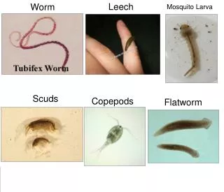
Treatment • Surgical removal of the worm from the anterior chamber under local anesthesia • Microscope examination • Dirofilaria tenuis ( 27 mm x 0.5 mm )
Fundus OD Multifocal choroiditis with RPE & chorioretinal scars in the periphery.
Investigations • Normal • Total & differential leucocyte count • Peripheral blood smear • Urine & stool test
Treatment • Diethyl carbamazine citrate (6mg/kg/day) given in two divided doses for 3 weeks.
Follow up • After 1 month • BCVA OD – 6/6 • Fundus • OD - Healed choroiditis • OS - Normal
Fundus OD
Conclusion • Live intraocular worm may present as acute iridocyclitis with multifocal choroiditis without any systemic complaints. • Surgical removal of the worm and antibiotic therapy results in resolution of choroiditis & improvement in visual acuity.