Download
1 / 20
200 likes | 374 Views
Energy generation in mitochondria II. Metals tightly bound to proteins from versatile electron carriers Protons are readily moved by transfer of electrons Electron transfers release large amounts of energy

E N D
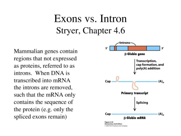
Energy generation in mitochondria II Metals tightly bound to proteins from versatile electron carriers Protons are readily moved by transfer of electrons Electron transfers release large amounts of energy Respiratory chain and ATP synthase are biochemically separate systems linked only by a proton-motive force Refer to chapter 18, Stryer, 5e Lecture 23, Michael Schweizer
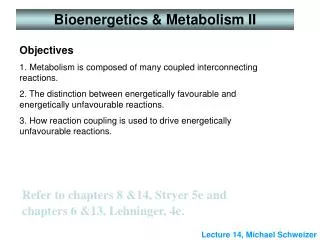
ElectronCarriers NAD+/NADH and FAD/FADH2 were introduced earlier. FMN (Flavin MonoNucleotide) is a prosthetic group of some flavoproteins. It is similar in structure to FAD (Flavin Adenine Dinucleotide), but lacking the adenine nucleotide. When free in solution, FMN (like FAD) can accept 2 e- + 2 H+ to form FMNH2.
Iron-sulfur Centers Iron-sulfur centers transfer only one electron, even if they contain two or more iron atoms, because of the close proximity of the iron atoms. e.g., a 4-Fe center might cycle between redox states: Fe+++3,Fe++1(oxidized)+1 e-Fe+++2, Fe++2(reduced)
Iron-sulfur Centers (A) A single iron ion bound by 4 cysteine residues • [2Fe-2S]; • (b) [4Fe-4S] Karp 3e, Figure 5.12
Cytochromes Cytochromesabsorb light at characteristic wavelengths. Absorbance changes upon oxidation/reduction of the heme iron provide a basis for monitoring redox state. Some cytochromes are part of large integral membrane complexes, each consisting of several polypeptides and multiple electron carriers. Cytochrome c is a small, water-soluble protein with a single heme group. Cytochrome c reversibly binds to integral membrane electron transfer complexes from which it receives, or to which it donates, an electron.
Hemes in the 3 classes of cytochrome (a,b,c) differ in substituents on the porphyrin ring.Only heme c is covalently linked to the protein via thioether bonds to Cys residues.
Cytochrome c receives electrons from complex III moves along outer surface of inner membrane donates electrons to complex IV Drawn by Dr B J Catley
Coenzyme Q (CoQ, Q or ubiquinone) is lipid-soluble. It dissolves in the hydrocarbon core of a membrane. Most often n = 10. The isoprene tail of Q10 is longer than the width of a lipid bilayer. CoQ has a quinone ring, which can be reduced to the quinol. Free CoQ can undergo a 2 e- oxidation/reduction: Q + 2 e- + 2 H+ QH2.
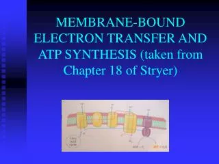
The generation of an H+ gradient across a membrane by electron transport reactions
pH 8 pH 7 The ATP-generating system of the inner mitochondrial membrane
Approximate positions on the redox potential scale of the electron transport complexes. The scale on the right gives the approximate free energy released when a pair of electrons is transferred from components to oxygen
Apparatus for measurement of redox potentials Karp 3e, Figure 5.10
The redox potential is a measure of electron affinities Examples of redox potentials: redox potential E0’ NADH NAD+ + H+ + 2 e- -320 mV QH2 red Qox + 2 H+ + 2 e- + 30 mV Cytred Cytox + e- +230 mV H2O ½ O2 + 2 H+ + 2 e- + 820 mV Difference in redox potential from NADH to O2= 1140 mV or 1.14 V; DGO’ = -26.2 kcal .mol-1 ( -52.4 kcal . mol-1 for 2 electrons transferred per NADH molecule)
Calculation ofDGO’ from redox potentials For an electron transfer: DE°' =E°'(oxidant) – E°'(reductant) = E°'(acceptor) – E°'(donor) DGo' = – nFDE°' (E°' is the mid-point potential) An electron transfer reaction is spontaneous (negative DG) if E°' of the donor is more negative than E°' of the acceptor, i.e., when there is a positive DE°‘.
ElectronTransfer Consider transfer of 2 electrons from NADH to oxygen: a. ½ O2 + 2H+ + 2e- H2O E°' = +0.820 V b. NAD+ + 2H+ + 2e- NADH + H+ E°' = -0.320 V Subtracting reaction b from a: c. ½ O2 + NADH + H+ H2O + NAD+ DE°'= +1.14 V DG = - nFDEo' = – 2(96494)(1.14) = (-220 kJ mol-1) = -52.4 kcal mol-1
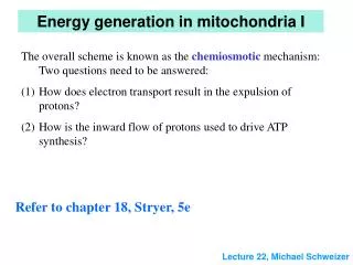
How are protons ejected? Generation of ATP in mitochondria by the chemiosmotic mechanism
The mechanism of proton translocation by complex III as a result of electron transport in mitochondria
Complex III QH2 + 2 H+ matrix + 2 cyt cox Q + 4 H+ cytosol + 2 cyt cred complex III contains 2 distinct binding sites for Q (see Figure 18.17, p593, Stryer 5e!); in 1 cycle 2 QH2 enter the pathway. Only one molecule of QH2 is oxidised, but achieves the ejection of 4 protons!
(a) Ubiquinone or coenzyme Q structure (b) Oxidised, semiquinone and reduced forms of Q
Schematic representation of the components of the electron transport chain within the inner mitochondrial membrane Karp, 3e, Figure 5.16