The Special Senses
450 likes | 488 Views
Explore the intricate workings of the special senses - smell, taste, vision, hearing, and equilibrium. Discover the anatomy and physiology of olfaction, gustation, and eye structures, enhancing your understanding of these vital sensory functions.

The Special Senses
E N D
Presentation Transcript
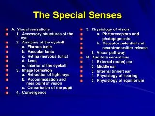
The Special Senses • Smell, taste, vision, hearing and equilibrium • Housed in complex sensory organs
Chemical Senses • Interaction of molecules with receptor cells • Olfaction (smell) and gustation (taste) • Both project to cerebral cortex & limbic system • evokes strong emotional reactions
Olfactory Epithelium • 1 square inch of membrane holding 10-100 million receptors • Covers superior nasal cavity and cribriform plate • 3 types of receptor cells
Cells of the Olfactory Membrane • Olfactory receptors • bipolar neurons with cilia or olfactory hairs • Supporting cells • columnar epithelium • Basal cells = stem cells • replace receptors monthly • Olfactory glands • produce mucus • Both epithelium & glands innervated cranial nerve VII (facial nerve)
Olfaction: Sense of Smell • Odorants bind to receptors • Na+ channels open • Depolarization occurs • Nerve impulse is triggered
Adaptation & Odor Thresholds • Adaptation = decreasing sensitivity • Olfactory adaptation is rapid • 50% in 1 second • complete in 1 minute • Low threshold • only a few molecules need to be present • methyl mercaptan added to natural gas as warning
Olfactory Pathway • Axons from olfactory receptors form the olfactory nerves (Cranial nerve I) that synapse in the olfactory bulb • pass through 40 foramina in cribriform plate • Neurons within the olfactory bulb form the olfactory tract that synapses on primary olfactory area of temporal lobe • conscious awareness of smell begins • Other pathways lead to the frontal lobe where identification of the odor occurs
Gustatory Sensation: Taste • Taste requires dissolving of substances • Five classes of stimuli--sour, bitter, sweet, salty, and umami (meaty or savory) • 10,000 taste buds found on tongue, soft palate & larynx • Found on sides of circumvallate & fungiform papillae • 3 cell types: supporting, receptor & basal cells
Anatomy of Taste Buds • An oval body consisting of 50 receptor cells surrounded by supporting cells • A single gustatory hair projects upward through the taste pore • Basal cells develop into new receptor cells every 10 days.
Physiology of Taste • Complete adaptation in 1 to 5 minutes • Thresholds for tastes vary among the 5 primary tastes • most sensitive to bitter (poisons) • least sensitive to salty and sweet • Mechanism • dissolved substance contacts gustatory hairs • receptor potential results in neurotransmitter release • nerve impulse formed neuron
Gustatory Pathway • Gustatory fibers found in cranial nerves • VII (facial) serves anterior 2/3 of tongue • IX (glossopharyngeal) serves posterior 1/3 of tongue • X (vagus) serves palate & epiglottis • Signals travel to thalamus or limbic system & hypothalamus • Taste fibers extend from the thalamus to the primary gustatory area on parietal lobe of the cerebral cortex • providing conscious perception of taste
Accessory Structures of Eye • Eyelids or palpebrae • protect & lubricate • epidermis, dermis, CT, orbicularis oculi m., tarsal plate, tarsal glands & conjunctiva • Tarsal glands • oily secretions keep lids from sticking together • Conjunctiva • palpebral (eyelids) & bulbar (sclera) • stops at corneal edge • dilated BV--bloodshot
Eyelashes & Eyebrows Eyeball = 1 inch diameter 5/6 of Eyeball inside orbit & protected • Eyelashes & eyebrows help protect from foreign objects, perspiration & sunlight • Sebaceous glands are found at base of eyelashes (sty) • Palpebral fissure is gap between the eyelids
Lacrimal Apparatus • About 1 ml of tears produced per day. Spread over eye by blinking. Contains bactericidal enzyme called lysozyme.
Extraocular Muscles • Six muscles that insert on the exterior surface of the eyeball • Innervated by CN III, IV or VI. • 4 rectus muscles -- superior, inferior, lateral and medial • 2 oblique muscles -- inferior and superior
Tunics (Layers) of Eyeball • Fibrous Tunic(outer layer, cornea and sclera) • Vascular Tunic (middle layer) • Nervous Tunic(inner layer)
Fibrous Tunic -- Description of Cornea • Transparent • Helps focus light (refraction) • astigmatism • 3 layers • nonkeratinized stratified squamous • collagen fibers & fibroblasts • simple squamous epithelium • Transplants • common & successful • no blood vessels so no antibodies to cause rejection • Nourished by tears & aqueous humor
Fibrous Tunic -- Description of Sclera • “White” of the eye • Dense irregular connective tissue layer -- collagen & fibroblasts • Provides shape & support • At the junction of the sclera and cornea is an opening (Canal of Schlemm) • Posteriorly pierced by Optic Nerve (CNII)
Vascular Tunic -- Choroid & Ciliary Body • Choroid • pigmented epithilial cells (melanocytes) & blood vessels • provides nutrients to retina • black pigment in melanocytes absorb scattered light • Ciliary body • ciliary processes • folds on ciliary body • secrete aqueous humor • ciliary muscle • smooth muscle that alters shape of lens
Vascular Tunic -- Iris & Pupil • Colored portion of eye • Contains melanin • Shape of flat donut suspended between cornea & lens • Hole in center is pupil • Function is to regulate amount of light entering eye • Autonomic reflexes • circular muscle fibers contract in bright light to shrink pupil • radial muscle fibers contract in dim light to enlarge pupil
Vascular Tunic -- Description of lens • Avascular • Crystallin proteins arranged like layers in onion • Clear capsule & perfectly transparent • Lens held in place by suspensory ligaments • Focuses light on fovea (back surface of eye)
Vascular Tunic -- Suspensory ligament • Suspensory ligaments attach lens to ciliary process • Ciliary muscle controls tension on ligaments & lens
Nervous Tunic -- Retina • Posterior 3/4 of eyeball • Optic disc • optic nerve exiting back of eyeball • Central retina BV • fan out to supply nourishment to retina • visible for inspection • hypertension & diabetes • Detached retina • trauma (boxing) • fluid between layers • distortion or blindness View with Ophthalmoscope
Layers of Retina • Pigmented epithelium • nonvisual portion • absorbs stray light & helps keep image clear • 3 layers of neurons (outgrowth of brain) • photoreceptor layer • bipolar neuron layer • ganglion neuron layer
Rods & Cones--Photoreceptors • Rods----rod shaped • shades of gray in dim light • 120 million rod cells • discriminates shapes & movements • distributed along periphery • Cones----cone shaped • sharp, color vision • 6 million • fovea of macula lutea • densely packed region • at exact visual axis of eye • 2nd cells do not cover cones • sharpest resolution or acuity
Pathway of Nerve Signal in Retina • Light penetrates retina • Rods & cones transduce light into action potentials • Rods & cones excite bipolar cells • Bipolars excite ganglion cells • Axons of ganglion cells form optic nerve leaving the eyeball (blind spot) • To thalamus & then the primary visual cortex
Cavities of the Interior of Eyeball • Anterior cavity (anterior to lens) • filled with aqueous humor • produced by ciliary body • continually drained • replaced every 90 minutes • 2 chambers • anterior chamber between cornea and iris • posterior chamber between iris and lens • Posterior cavity (posterior to lens) • filled with vitreous body (jellylike) • formed once during embryonic life • floaters are debris in vitreous of older individuals
Aqueous Humor • Continuously produced by ciliary body • Flows from posterior chamberinto anterior through the pupil • Scleral venous sinus • canal of Schlemm • opening in white of eyeat junction of cornea & sclera • drainage of aqueous humor from eye to bloodstream • Glaucoma • increased intraocular pressure that could produce blindness • problem with drainage of aqueous humor
Major Processes of Image Formation • Refraction of light • by cornea & lens • light rays must fall upon the retina • Accommodation of the lens • changing shape of lens so that light is focused • Constriction of the pupil • less light enters the eye
External Ear • Function = collect sounds • Structures • auricle or pinna • elastic cartilage covered with skin • external auditory canal • curved 1” tube of cartilage & bone leading into temporal bone • ceruminous glands produce cerumen = ear wax • tympanic membrane or eardrum • epidermis, collagen & elastic fibers, simple cuboidal epith. • Perforated eardrum (hole is present) • at time of injury (pain, ringing, hearing loss, dizziness) • caused by explosion, scuba diving, or ear infection
Middle Ear Cavity • Air-filled cavity in the temporal bone • Separated from external ear by eardrum and from internal ear by oval & round window • 3 ear ossicles connected by synovial joints • malleus attached to eardrum, incus, stapes attached to membrane of oval window • Auditory tube leads to nasopharynx • helps to equalize pressure on both sides of eardrum
Inner Ear---Bony Labyrinth Vestibule canals • Bony labyrinth = set of tubelike cavities in temporal bone • semicircular canals, vestibule & cochlea lined with periosteum & filled with perilymph • surrounds & protects Membranous Labyrinth ampulla
Inner Ear---Membranous Labyrinth • Membranous labyrinth = set of membranous tubes containing sensory receptors for hearing & balance and filled with endolymph • utricle, saccule, ampulla, 3 semicircular ducts & cochlea
Cranial nerves of the Ear Region • Vestibulocochlear nerve = CN VIII
Cochlear Anatomy • 3 fluid filled channels found within the cochlea • scala vestibuli, scala tympani and cochlear duct • Vibration of the stapes upon the oval window sends vibrations into the fluid of the scala vestibuli
Tubular Structures of the Cochlea • Stapes pushes on fluid of scala vestibuli at oval window • At helicotrema, vibration moves into scala tympani • Fluid vibration dissipated at round window which bulges • The central structure is vibrated (cochlear duct)
Section thru one turn of Cochlea • Partitions that separate the channels are Y shaped • vestibular membrane above & basilar membrane below form the central fluid filled chamber (cochlear duct) • Fluid vibrations affect hair cells in cochlear duct
Anatomy of the Organ of Corti • 16,000 hair cells have 30-100 stereocilia (microvilli) • Microvilli make contact with tectorial membrane (gelatinous membrane that overlaps the spiral organ of Corti) • Basal sides of inner hair cells synapse with 1st order sensory neurons whose cell body is in spiral ganglion
Physiology of Hearing • Auricle collects sound waves • Eardrum vibrates • slow vibration in response to low-pitched sounds • rapid vibration in response to high-pitched sounds • Ossicles vibrate since malleus attached to eardrum • Stapes pushes on oval window producing fluid pressure waves in scala vestibuli & tympani • oval window vibration 20X more vigorous than eardrum • Pressure fluctuations inside cochlear duct move the hair cells against the tectorial membrane • Microvilli are bent producing receptor potentials
Physiology of Equilibrium (Balance) • Static equilibrium • maintain the position of the body (head) relative to the force of gravity • macula receptors within saccule & utricle • Dynamic equilibrium • maintain body position (head) during sudden movement of any type--rotation, deceleration or acceleration • crista receptors within ampulla of semicircular ducts
Vestibular Apparatus • Notice: semicircular ducts with ampulla, utricle & saccule