Download
1 / 21
220 likes | 809 Views
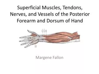
Extensor Side of the forearm and Hand. Phil Pullen, D.O. Garden City Hospital Orthopaedic Surgery Resident. Extensor muscles of the forearm. Can be divided into 3 functional groups:

E N D
Extensor Side of the forearm and Hand Phil Pullen, D.O. Garden City Hospital Orthopaedic Surgery Resident
Extensor muscles of the forearm • Can be divided into 3 functional groups: • Muscles that extend and abduct or adduct the hand (extensor carpi radialis longus, extensor carpi radialis brevis, and ext. carpi ulnaris) • Muscles that extend the medial 4 digits (ext digitorum, ext indicis, and ext digiti minimi) • Muscles that extend or abduct the thumb (abductor pollicis longus, ext pollicis brevis, and ext pollicis longus)
Muscles • Brachioradialis: • origin – lateral supracondylar ridge of the humerus • Insertion- lateral surface of distal end of radius • Innervation – radial nerve • Action –flexes the forearm • Extensor Carpi Radialis Longus: • Origin – lateral supracondylar ridge • Insertion-base of 2nd MC • Innervation- radial nerve • Action- extend and abduct at wrist
Muscles • Extensor Carpi Radialis Brevis: • Origin – lateral epicondyle of humerus • Insertion- base of 3rd MC • Innervation – deep branch of radial n. • Action – extend and abduct at wrist • Extensor Digitorum • Origin – lateral epicondyle • Insertion – extensor expansions of medial four digits • Innervation – posterior interosseus • Action – extends medial 4 digits at MCP and extends wrist
Muscles • Extensor Digiti Minimi • Origin – lateral epicondyle • Insertion – extensor expansion of 5th digit • Innervation – posterior interosseus • Action – extends 5th digit at MCP and IP joints • Extensor Carpi Ulnaris • Origin – lateral epicondyle • Insertion – base of 5th MC • Innervation – post interosseus • Action – extends and adducts at wrist
Muscles • Supinator: • Origin – lateral epicondyle and posterior border of the ulna • Insertion – lateral, post., and ant. Surfaces of the prox. 3rd of the radius • Innervation – deep branch of the radial • Action- supinates the forearm
Muscles • Abductor Pollicis Longus: • Origin – post surfaces of ulna, radius and interosseus membrane • Insertion – base of 1st MC • Innervation – post interosseus • Action – abducts thumb and extends at the CMC joint
Muscles • Extensor Pollicis Brevis • Origin – post surface of the radius and interosseus membrane • Insertion – base of prox phalanx of thumb • Innervation – post interosseus • Action – extends prox phalanx of thumb at CMC joint • Extensor Pollicis Longus • Origin – post surface of middle 3rd of ulna and interosseus membrane • Insertion – base of distal phalanx of thumb • Innervation – post interosseus • Action – extends distal phalanx of thumb at MCP and IP joints
Muscles • Extensor Indicis • Origin – post surface of ulna and interosseus membrane • Insertion – extensor expansion of 2nd digit • Innervation – post interosseus • Action – extends 2nd digit and helps to extend hand
Extensor mechanism • At the distal end of the MC and along the phalanges, the ext tendons flatten out to form the extensor expansions • Each ext digital expansion is a triangular tendinous aponeurosis that wraps around the dorsum of the head of the MC and prox phalanx
Extensor mechanism • The hood is anchored on both sides to the palmar ligament to hold the tendon in the middle of the digit • This expansion divides into a median band that passes to the base of the middle phalanx • And 2 lateral bands that pass to the base of the distal phalanx
Extensor mechanism • The interosseus and lumbrical muscles attach to the lateral bands of the extensor expansion
Extensor Compartments • I – abductor pollicis longus, extensor pollicis brevis • II – extensor carpi radialis longus and brevis • III – extensor pollicis longus • IV – extensor digitorum, extensor indicis • V – extensor digiti minimi • VI – extensor carpi ulnaris
Nerves • Radial nerve leaves the posterior compartment of the arm to cross the anterior aspect of the lateral epicondyle • After it enters the forearm it branches into the deep and superficial branches and the posterior cutaneous nerve of the forearm • Nerve supply to the extensor muscles includes the radial nerve, the deep branch of the radial nerve and the posterior interosseus nerve which is a continuation of the deep branch of the radial nerve
Arterial Supply to Extensor Side of Forearm • The arterial supply to the extensors of the forearm come from branches from the ulnar artery, namely the posterior interosseus artery with its muscular branches • The ulnar artery (the larger of two branches of the brachial artery) begins in the cubital fossa just medial to the biceps tendon