
Mechanical Ventilators
E N D
Presentation Transcript
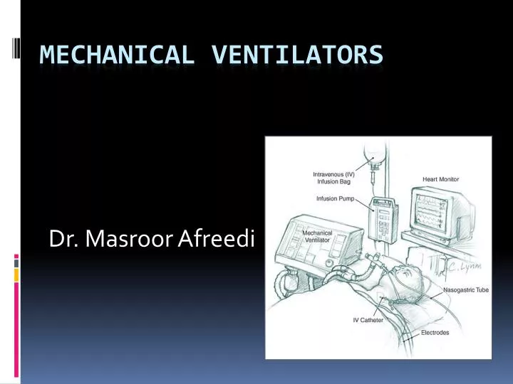
Dr. Masroor Afreedi Mechanical Ventilators
Early History of Ancient Times • Ancient writings by the Egyptians and Greeks described theories of respiration. • In the Old testament there is a mention of Prophet Elisha inducing pressure breathing from his mouth into the mouth of a child who was dying–(Kings 4:34-35). Hippocrates (460-375 BC) wrote the first description of endotracheal intubation his book –‘Treatise on Air’ “One should introduce a cannula into the trachea along the jaw bone so that air can be drawn into the lungs”.
Modern History • Paracelsus (1493-1541) used ‘Fire Bellows’ connected to a tube inserted into patient’s mouth as a device for assisted ventilation. This was the first study (1550) which credited him with the first form of mechanical ventilation. • Vesalius (1543) performed ventilation via a tracheostomy in a pig. • Hook (1667) used bellows via a tracheostomy in a dog. • John Fathergill in 1744 reported a successful case of ‘mouth to mouth’ resuscitation.
Modern History John Hunter developed double bellows for resuscitation in 1775 - one for blowing air in and the other for drawing bad air out
Origins of mechanical ventilation The era of intensive care medicine began with positive-pressure ventilation • Negative-pressure ventilators (“iron lungs”) • Non-invasive ventilation first used in Boston Children’s Hospital in 1928 • Used extensively during polio outbreaks in 1940s – 1950s • Positive-pressure ventilators • Invasive ventilation first used at Massachusetts General Hospital in 1955 • Now the modern standard of mechanical ventilation The iron lung created negative pressure in abdomen as well as the chest, decreasing cardiac output. Iron lung polio ward at Rancho Los Amigos Hospital in 1953.
Negative Pressure Ventilators From the mid 1800-1900s a large number of devices were invented that applied negative pressure around the body or thoracic cavity – these devices became known as negative pressure ventilators or 'iron lungs'. Two successful designs became popular; in one, the body of the patient was enclosed in an iron box or cylinder and the patient’s head protruded out of the end. The second design was a box or shell that fitted over the thoracic area only (chest cuirass). Patients with chronic paralytic disorders were successfully ventilated on this cuirass ventilators at home for 25-30 years
Scandinavian Polio Epidemic - 1952 Between July-December of 1952, in Copenhagen, 2722 patients with poliomyelitis were treated in the Community Disease Hospital of which 315 patients required ventilatory support. Many principles of IPPV were defined during that time –including the use of cuffed tubes, periodic sigh breaths and weaning by reduction of assisted breaths. Towards the end of the epidemic a few positive pressure ventilators were invented (the Engstrom, Lundi and the Bang) which became popularly known as mechanical students.
Era of Respiratory Intensive Care • After polio epidemics, the 1960’s became an era of respiratory intensive care. Positive pressure ventilation with use of an artificial airway replaced the bulky and cumbersome negative pressure technology of respiratory support.
Era of Respiratory Intensive Care • Two types of ventilators and two modes of mechanical ventilation evolved during this period; • the first type of ventilator was pressure cycled (PCV). Two ventilators were commonly used for PCV in the 1960’s and 1970’s; the Bird Mark 7 and the Bennet PR2.
Era of Respiratory Intensive Care • The second type of ventilator that evolved from a historical perspective is the volume cycled ventilator – VCV. • The first fluidic ventilator utilizing moving streams of liquid or gas for sensing, logic, amplification and controls was designed for the US army in 1964 by Barila and • the first commercial versatile fluidic ventilator “Hamilton standard PAD” appeared in 1970. The term ‘weaning’ was used to explain various techniques to test the quality of patient’s spontaneous ventilation before extubation.
Present day • A mechanical change of substantial importance in the late 1960’s and early 1970’s that shaped the present era was the introduction of Positive End Expiratory Pressure (PEEP). • Two modes of ventilation Assisted Ventilation (AV) and Controlled Mechanical Ventilation (CMV) came together in a single piece of equipment and the modern era of multiple choice respiratory support was born. • The introduction of IMV permitted spontaneous respiration in the midst of substantial respiratory failure which paved the way for a means of weaning i.e. SIMV. PSV proved to be an addition to IMV that facilitated spontaneously breathing patients
INDICATIONS FOR MV • Hypoxemia • Acute respiratory acidosis • Reverse ventilatory muscle fatigue • Permit sedation and/or neuromuscular blockade • Decrease systemic or myocardial oxygen consumption
INDICATIONS CONTINUED • Reduce intracranial pressure through controlled hyperventilation • Stabilize the chest wall • Protect airway • Neurologic impairment • airway obstruction
TYPES OF CONVENTIONAL MV • Timed cycled • Home ventilators • Pressure cycled • Pressure controlled • Volume cycled • Flow cycled • Pressure support
VOLUME VENTILATION • Controlled mechanical ventilation CMV • Assist-control AC • Synchronized intermittent mandatory ventilation SIMV • Which mode?
VENTILATOR SETTINGS • Tidal volume • 10 to 15 mL/kg • Respiratory rate • 10 to 20 breaths/minute • normal minute ventilation 4 to 6 L/min • Fraction of inspired oxygen • Flow rate and I:E ratio
PRESSURE SUPPORT VENTILATION • Flow cycled • preset pressure sustained until inspiratory flow tapers to 25% of maximal value • Comfortable • Used mainly as a weaning mode • Wean pressure until equivalent to air way resistance • peak - plateau pressure
PRESSURE CONTROLED VENTILATION • Pressure cycled • Volume varies with lung mechanics • Minute ventilation is not assured • Improves oxygenation • recruitment of alveoli • Lessens volutrauma?
SETTINGS FOR PRESSURE CONTROL VENTILATION • Inspiratory pressure • I:E ratio • 1:2, 1:1, 2:1, 3:1 • Rate • FIO2 • Peep
PRESSURE REGULATED VOLUME CONTROLLED • Ventilate with pressure control • Preset volume • Inspiratory pressure is adjusted breath to breath • Minute ventilation is maintained
INDICATIONS FOR PEEP • ARDS • Stabilize chest wall • Physiologic peep • Decrease Auto-peep?
CONTRAINDICATIONS FOR PEEP • Increased intracranial pressure • Unilateral pneumonia • Bronchoplueral fistulae
PEEP • Increases FRC • Recruits alveoli • Improves oxygenation • Best Peep • based on lower inflection of pressure volume curve
TROUBLE SHOOTING VOLUME VENTILATION • High pressure alarm • Breath sounds • CXR • Low tidal volume • disconnected • Desaturation
TROUBLE SHOOTING PRESSURE VENTILATION • Low tidal volumes or minute ventilation • Desaturation • Breath sounds • Patient agitation • CXR
Sedation in Mechanically Ventilated Patients • Benzodiazepines • Opioids • Neuroleptics • Propofol • Ketamine • Dexmedetomidine
Benzodiazepines • Lorazepam • Half-life 12 to 15 hours • Major metabolite inactive • Midazolam • Half-life 1-4 hours, increased in cirrhosis, CHF, obesity, elderly • Active metabolite
Opioid • Morphine • Fentanyl • Hydromorphone
Neuroleptics • Haloperidol • Mild agitation .5mg to 2mg • Moderate agitation 2 to 5 mg • Severe 10 to 20 mg • Side Effects • Acute dystonic reactions • Polymorphic VT • Neuroleptic malignant syndrome
Propofol • Side Effect • Hypotension • Bradycardia • Anticonvulsant • Expensive • Use short term
Ketamine • Dissociative anesthetic state • Direct cardiovascular stimulant • Brochodilator • Side Effects • Dysphoric reactions • increased ICP
Dexmedetomidine • Centrally acting alpha 2 agonist • Approved for 24 hours or less • Side Effects • Hypotension • Bradycardia • Atrial fibrillation
Maintenance of Sedation • Titrate dose to ordered scale • Motor Activity Assessment Scale MAAS • Sedation-Agitation Scale SAS • Ramsay • Rebolus prior to all increases in the maintenance infusion • Daily interruption of sedation
NEUROMUSCULAR BLOCKING AGENTS • Difficult to asses adequacy of sedation • Polyneuropathy of the critically ill • Use if unable to ventilate patient after patient adequately sedated • Have no sedative or analgesic properties
Neuromuscular Blocking Agents • Depolarizing • Bind to cholinergic receptors on the motor endplate • Nondepolarizing • Competitively inhibit Ach receptor on the motor endplate
Depolarizing NMBASuccinylcholine • Rapid onset less than 1 minute • Duration of action is 7-8 minutes • Pseudocholinesterase deficiency • 1 in 3200 • Side Effects • Hyperthermia, Hyperkalemia, arrhythmias • Increased ICP
Nondepolarizing Agents • Pancuronium • Drug of choice for normal hepatic and renal function • Atracurium or Cisatracurium • Use in patients with hepatic and/or renal insufficiency • Vecuronium • Drug of choice for cardiovascular instability
No bubble is so iridescent or floats longer than that blown by the successful teacher.Sir William Osler
MV IN OBTRUCTIVE AIRWAY DISEASE • Decrease barotrauma • related to mean airway pressure • Increase I:E • decrease TV and/or increase flow • Minimize auto-peep • auto-peep shown to cause most barotrauma • Permissive hypercapnea
ARDS • Set peep to pressure shown at lower inflection point of pressure volume curve • Tidal volumes set below upper inflection point of pressure volume curve • Use pressure control ventilation early • Minimize volutrauma
Ventilation With Lower Tidal Volumes • Tidal volume: 6 ml/kg • Male 50 + 0.91(centimeters of height-152.4) • Female 45.5+0.91(centimeters of ht - 152.4) • Decrease or Increase TV by 1ml/kg to maintain plateau pressure 25 to 30. • Minimum TV 4ml/kg • PaO2 55 - 88 mm Hg. Sats 88 to 95% • pH 7.3 to 7.45
CASE EXAMPLE • 34 y/o female admitted with status asthmaticus and respiratory failure • You are called to see patient for inability to ventilate • Tidal volume 800 cc, FIO2 100%, AC 12 Peep 5 cm • PAP 70, returned TV 200 cc
Case example continued • Examine patient • CXR • Sedate • Assess auto-peep • Increase I:E • Lower PAP and MAP • Reverse bronchospasm & elect. Hypovent.
CONCLUSION • Three options for ventilation • volume, pressure, flow • Peep, know when to say no • Always assess to prevent barotrauma • ventilate below upper inflection point • assess static compliance daily • monitor for auto-peep