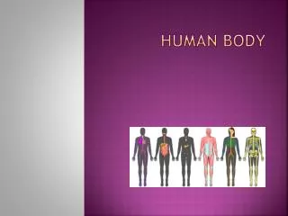
HUMAN BODY
HUMAN BODY. I. The Human Body. A. Introduction Humans are the most complex organisms on Earth, and every cell in the human body must work together to keep functioning. There are many levels of organization:

HUMAN BODY
E N D
Presentation Transcript
I. The Human Body A. Introduction Humans are the most complex organisms on Earth, and every cell in the human body must work togetherto keep functioning. There are many levels of organization: • 1. Cell - basic unit of structure and function in ALL living things and usually are specialized for a particular function. • 2. Tissues- group of cells that perform a singlefunction.
a. Epithelial • covers body surfaces, lines cavities, organs, vessels. May contain glands for secretions or cells with cilia. Examples include thyroid gland, epicardium of heart, arteries & veins. Epidermissmall intestine kidney
b. Connective • the most abundant and widespread tissue in the body. Used for support, transport, storage, and as connectors. Has a network of non-living material called a matrix. Ex. Bone, blood, cartilage.
c. Muscle • able to generate electrical signals that create force and movement. Ex. Skeletal muscle, cardiac muscle, & smooth muscle.
d. Nerve • specialized to generate and transmit electrical signals to transfer information. Examples include tissues of the brain and spinal cord. PNS – ganglion Blue stained Myelin sheath with red nuceli
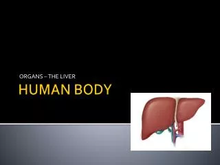
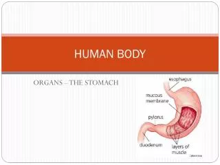
3. Organ - group of tissues working together for a particular function. 4. Organ system - Group of organs working together to perform a function.
B. Organ Systems • All the parts of the body work together to maintain the internal conditions inside of a human. 1. Nervous system - Receives, processes, and responds to sensory information: coordinates all body functions 2. Endocrine System - regulates homeostasis with chemicals called hormones. 3. Skeletal System -protects and supportsbody parts. 4. Muscular System - produces movement. 5.Integumentary System- physical barrieragainst injury, dehydration, pathogens.
6. Circulatory system - transports O2 and CO2, nutrients, and wastes. 7. Respiratory system - responsible for the exchangeof O2 and Co2 8. Lymphatic System - destroys pathogens, fights infection. 9. Digestive System -breaks downfood into absorbable monomers. 10. Urinary System - washes or cleans the blood; regulates blood volume. 11. Reproductive System – produces gametes; development of embryo in female
Organ systems work together in the body to maintain homeostasis. • Homeostasis - the process by which organisms keep internal and physical conditions constant, or in internal harmony. This harmony is accomplished by negative feedback mechanisms. • Negative feedback - the process in which a stimulus produces a response that opposesthe original stimulus. (ex. House hold heater or AC(pg. 896) & thyroid hormones) • This process is fulfilled by the endocrine and nervous systems working together. • Endocrine system - may take hours to couple of daysto affect the body. • Nervous system - quick and rapidtransmission to the body
Nervous System “I think, therefore, I am”
II. Nervous System • The nervous system controls and coordinates functions throughout the body, and responds to internal and external stimuli.
A. Neurons • Neurons are cells that transmitnerve impulses. Impulses are electrical signals. These signals contain information of what is happening to the body, what is going on in the body, and how the body should respond.
2. Structure of a Neuron • All neurons have the samebasicstructure.
Structure of a Neuron cont. • Cell Body - contains the nucleus and the rest of the organelles. Site of metabolic activity; it receives the impulses from the dendrites.
Structure of a Neuron cont. b. Dendrites - short, branched extensions that receivea stimulus and carry that information to the cell body; they act as detectors or antennae.
Structure of a Neuron cont. c. Axon - a long, single fiber that branches into many swellings known as axon terminals. The axon transmits impulses away from the cell body and out through the terminals.
2. Structure of a Neuron d. Schwann cells - specialized cells that coverthe axons of many larger neurons. The phospholipid bilayer of the cell membranes of these cells have a higher than usual concentration of lipids. Schwann cellswrap around the neuron to form insulation layers known as the Myelin sheath,which protects and insulates the neuron.
e. Nodes of Ranvier - nodes are small gaps in the myelin sheath. The electrical impulses can hopfrom node to node, allowing the impulse to travel faster and in one direction.
3. Synapse • The small space betweena neuron and any other cell is called the synapse. When an impulse reaches the end of an axon, the impulse must be carried across the synapse by chemicals called neurotransmitters.
Neurotransmitters are stored in vesiclesin the axon terminals of a neuron. When an impulse reaches the axon terminal, the vesicles containing the neurotransmitters are released into the synapse by exocytosis.
The chemicals diffuse across the synapse and stimulate the sodium channels to open in the adjacent cell, stimulating the next cell to go from resting potential to action potential. The neurotransmitters are then broken down and recycled. Examples of Neurotransmitters are Dopamine, Serotonin, Acetylcholine
4. All or Nothing response • The transmission of a nerve impulse is an all or nothing response which means that either the stimulus will produce an impulse or it will not produce an impulse; therefore the strength of an impulse is always the same. The minimumlevelof a stimulus required to activate a neuron called the threshold. Because all nerve impulses have the same intensity, the intensity of the stimulus (or response) is reflected in the number of neurons stimulated and the frequencyof stimulation.
III. Organization of the Human Nervous System A. Central Nervous System (CNS) The CNS is made up of the brain and the spinal cord.
A. Central Nervous System (CNS) 1. Protection of the CNS - These organs are vital importance and are therefore, well protected. Three means of protection: a. Bones - Cranium protects the brain; vertebrae protect the spinal cord b. Meninges - 3 layers of tough, elastic connective tissue that cushion the brain and spinal cord. c. Cerebrospinal Fluid - fluid between the meninges; acts as additional cushion
2. Spinal Cord • Column of nerve tissue extending from the brain through the vertebrae. Serves as the main communication linkbetween the brain and the rest of the body
3. Brain • It is the control centerof the entire nervous system. Although it makes up only about 2% of the body’s mass, it uses about 25% of the body’s oxygen. The human brain is folded into convolutions to increase surface area.
Parts of the brain: a. Cerebrum b. Cerebellum c. Brain Stem d. Thalamus e. Hypothalamus
a. Cerebrum 1.Cerebrum- Largest part of the brain; it is composed of 2hemispheres or sides - right & left. It controls the conscious activities, intelligence, memory, language, sense, judgment, movement, emotions, personality
Corpus Calleosum - connects the right and left cerebral hemispheres and maintains the communication between them.
b. Cerebellum • located in the back of the brain. Regulates posture, balance, muscle tone, & coordinates the voluntary movement.
c. Brain Stem Composed of the medulla oblongata, pons, & midbrain. 1 & 2. Pons & midbrain - act as pathways that connect the different parts of the brain. 3. Medulla oblongata - controls involuntary activities such as breathing, heart rate, and blood pressure
d. Thalamus • “telephone operator” for the brain it is the main site of sensory processing; sorts out sensory information – ex: smell, taste, etc.
e. Hypothalamus • Controls many activities relating to homeostasis such as body temp, breathing rate, feeling of hunger, thirst, sex drive. Works closely with medulla oblongata.
B. Peripheral Nervous System (PNS) The PNS relays messages from the body to the brain and transmits commands from the brain to the body. The PNS is composed of nerves which are bundles of sensory and motor neurons. The PNS is subdivided into: • Autonomic Nervous System • Somatic Nervous System
1. Autonomic Nervous System • Network of nerves that regulate activities not under conscious control. The ANS is under the control of the medulla oblongata.
2. Somatic Nervous System • Regulates activities under conscious control, such as the movement of skeletal muscles. Sometimes a stimulus will result in an automatic, unconscious response within the somatic system. This is called a reflex. Most impulses received by the spinal cord are sent to the brain for processing and interpretation. A reflex is processed directly in the spinal cord. It is a quick and automaticresponse, although the brain becomes aware after the reflex has occurred.
IV. Sensory Organs • A. Vision • B. Hearing • C. Smelling and Taste • D. Touch & related senses
A. Vision • The eyes contain sensory receptors that respond to light. The light energy is then converted to an electrical impulseand transmitted to the brain.
A. Vision - cont 1. Light first passes through the cornea - transparent, protective covering which begins to focus the light onto the back of the eye. The light then enters a chamber filled with watery fluid called the aqueous humor.
2. The amount of lightentering eye is controlled by the iris which consists of tiny muscles arranged in a ring. 3.Light enters through the opening in the center of the iris called the pupil.
4. The beam of light passes through the lens. There are small muscles attached to the lens to change its shape and complete the focusing of the light. There is a chamber behind the lens filled with a jellylike fluidcall the vitreous humor.
A. Vision- cont 5. The lens focuses the light onto a delicate tissue called the retina where the light energy is converted to an electrical impulse. The retina is nerve tissue made up of 2 types of cells: A. rods - cells that detect shape and movement; are stimulated even in dimlight. B. cones - cells that detect color; require bright light for stimulation.
6. The retinal tissue comes together to form the optic nerve which transmits the impulse to the thalamus and then to the cerebrum 7. Sclera - the “white of the eye” and is a continuation of the cornea. It encases and protects the eye 8. Choroid - a layer of blood vesselsthat nourish the eye.