Download
1 / 19
190 likes | 418 Views
Cells and Development. Domains of Life. Current classification scheme has the largest division into 3 groups: Bacteria, Archaea, and Eukarya. Based on 16S ribosomal RNA sequence similarities. Bacteria (also called Eubacteria) and Archaea are prokaryotes. Eukarya are eukaryotes.

E N D
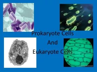
Domains of Life • Current classification scheme has the largest division into 3 groups: Bacteria, Archaea, and Eukarya. Based on 16S ribosomal RNA sequence similarities. • Bacteria (also called Eubacteria) and Archaea are prokaryotes. Eukarya are eukaryotes. • Prokaryote: no membrane-bound nucleus containing the cell’s DNA. Eukaryotes are defined by having a membrane-bound nucleus that holds the DNA. • Eukaryotes also have other membrane bound organelles, and eukaryotes have linear chromosomes. • Prokaryotes don’t have membrane bound organelles, although some prokaryotes have some internal membranes. Most prokaryotes have circular chromosomes, but some have linear chromosomes or even a mixture of circular an linear chromosomes.
Eukaryotic Cell Structures • nucleus: holds the chromosomes, surrounded by the double membrane nuclear envelope, which has nuclear pores in it--traffic is controlled, but ribosomes (big) can get out for example. The nucleolus is an area of the nucleus where ribosomal RNA is made in large quantities. Other structures in the nucleus have also been defined, including area for transcription and for RNA processing. • mitochondria: makes most of the ATP by aerobic respiration: Krebs cycle and electron transport/oxidative phosphorylation. Two membranes separate 2 different regions of the mitochondria. Mitochondria have their own circular DNA with about 30 genes on it: derived from bacterial DNA (endosymbiont hypothesis).
Eukaryotic Cell Structures • endoplasmic reticulum (ER): series of membrane-bound channels and vesicles in the cell. The rough ER is studded with ribosomes: for translation of proteins that get secreted or get inserted into the cell membrane. The smooth ER is where sugars are added to the proteins (glycosylation); membrane lipids are also synthesized in the smooth ER. • Golgi apparatus: takes proteins from the ER and packages them for secretion from the cell. Movement between the ER, the Golgi, and the plasma membrane occurs in small membrane-bound bodies called vesicles. • Lysosomes: intracellular digestion. Low internal pH and full of various hydrolytic enzymes. Vesicles full of extracellular material get transported from plasma membrane to the lysosomes; also involved in apoptosis (programmed cell death). • Peroxisomes: use superoxide and peroxide (very toxic to the cell) to oxidize various compounds.
Eukaryotic Cell Structures • Plasma membrane: the outer surface of the cell. Composed of a phospholipid bilayer: by itself only lets oxygen, carbon dioxide, water, as few other small molecules in or out. All other molecules are transported down the electrochemical gradient by channel proteins, or pumped up the gradient by ATP-driven pumps. Also, plasma membrane has adhesion proteins that connect to other cells or extracellular matrix, and receptors for hormones and other signaling molecules. • Cytoskeleton: microtubules, microfilaments, intermediate filaments: structure and transport within the cell. • microfilaments are made of actin, which interacts with various forms of myosin to provide cell movement and changes in cell shape. • microtubules are made of tubulin monomers. Mitotic spindle is made of microtubules. Also movement of organelles occurs with the interaction of microtubules and motor proteins. Cilia and flagella are made of microtubules as well. • intermediate filaments are structural only.
Eukaryotic Cells • Size is limited by the need for oxygen, nutrients, control signals, etc., to diffuse from one end of the cell to the other. For more-or-less round cells, 10-30 µm is typical. Most bacterial cells are 1-2 µm. • Many different cell types in humans. All have same basic DNA, with minor changes due to random mutations plus a few cell types (e.g. immune system cells) have major DNA changes. • Most human cells are diploid (2n). However: • the gametes, sperm and egg, are haploid (1n). • some are polyploid due to endomitosis, DNA replication without cell division. Examples: hepatocytes (liver cells) range from 2n to 8n, and bone marrow megakaryocytes range from 16n to 64n. • some cells, notably red blood cells, have no nucleus. • some cells fuse together to form a multinuclear syncytium, notably muscle cells. • In most animals, the germ line cells, cells which become the sperm and egg, are separated from the somatic cells (all other cells) early in development.
Cell Culture • How to study cell behavior. Can do it in whole organisms, sometimes called in situ studies. This can have lots of complicating factors as many tissues and organs interact. Also, can’t see or access many cells. • Tissue explants: cut out a piece, culture it in a nutrient medium • Primary cell culture: dissociate a tissue into individual cells and grow in nutrient medium. Problem: cells are mortal, after about 60 divisions they stop dividing. • Permanent cell line: Easy to grow and maintain. No limit to cell divisions, immortalized (transformed) by mutations equivalent to cancer induction. Can be done spontaneously, or with known mutagens, or by transfection with DNA containing oncogenes (mutated genes which cause cancer). • an important resource is cell lines from interesting patients. To keep a stable source of their genetic material, lymphoblastoid cells can be immortalized by Epstein-Barr virus, which remains separate from the chromosomal DNA.
Principles of Development • How to get from a single cell, the zygote, to a multicellular organism. • General events: • cell division and growth • differentiation: cells develop different phenotypes • pattern formation: overall development of body axes, the general body plant, and structure of individual organs • morphogenesis: changes in shape
Canalization of Development • Cells become increasingly specialized during development. Their range of possible fates (final cell type) decreases. This is called the canalization of development. • Initially, cells of the embryo are totipotent: can develop into any embryonic cell. After a while, embryo is divided into a trophoblast and an inner cell mass. Inner cell mass become the embryo while trophoblast becomes outer membranes and placenta. Cells in ICM can become any embryonic tissue, but they can’t become trophoblast cells: these cells are pluripotent. As development proceeds, embryonic cells become increasingly specialized and can no longer become any final cell type: they become multipotent and finally unipotent when they can only become one final cell type. • At some point, a cell is determined to be a particular cell type. Determination is followed by differentiation, changes of form and function, into that cell type. • decisions about determination are caused by a cell’s lineage: previous decisions, by its position in the embryo, and by signals passed between the cells.
Stem Cells • Stem cells are self-renewing cells that differentiate into a variety of cell types. After a stem cell divides, one daughter cell typically remains a stem cell, while the other one starts to differentiate into a final cell type: this is called asymmetric cell division. • There are many types of stem cell in adults, and they are generally rare and hard to find. Some differentiate into a single cell type, while others can have multiple fates. • Embryonic stem cells are the pluripotent cells of the inner cell mass, which can become any kind of embryonic cell.
Pattern Formation • How does the basic body plan get formed • axes: dorsal-ventral (back-front), cranial-caudal (head-tail), left-right. • position within an organ: e.g. how to get 5 different fingers on a hand • axis development: based partly an uneven distribution of components in the egg and partly on external events. • sperm entry point determines boundary between trophoblast and inner cell mass, which in turn determines the dorsal-ventral axis • cranial-caudal axis probably determined by position of second polar body exit relative to sperm entry point • morphogen gradients. Certain cells secrete chemicals that act as morphogens: signals that allow other cells in that tissue to determine their position in the tissue. The farther a cell is from the morphogen secretion site, the lower the concentration of the morphogen. • a well known example is the zone of polarizing activity, which occurs in limb buds. Cells nearest the ZPA become the little finger or toe, while those farthest away become the thumb or big toe.
Hox genes • Animal development along an axis, from Drosophila to humans, is largely determined by clusters of homeobox (Hox) genes. • Different members of the Hox clusters are activated in different parts of the morphogen gradient, in an overlapping pattern • The Hox gene products then stimulate the activity of other genes that cause the cells to differentiate into the proper type.
Fertilization • Egg is surrounded by two layers of extracellular matrix, the vitelline membrane and the zona pellucida. The sperm cells must dissolve their way through these layers to get to the egg. Sperm contain an acrosome at their tips that contains the necessary enzymes. • When a sperm reaches the egg membrane, the membranes fuse, putting teh sperm nucleus inside the egg. • Egg membrane then depolarizes and cortical granules release their contests to push all other sperm cells away. • Meiosis 2 occurs and the second polar body exits opposite the sperm entry point. • the male and female pronuclei then undergo mitosis together, and the resulting nuclei fuse. • Fertilization occurs in the Fallopian tubes. After fertilization, the embryo takes about a week to reach the uterus.
Early Development • The early cell divisions of the embryo occur without any overall growth. These divisions, the cleavage divisions, result in the morula, a ball of 16 or more cells. Each cell is called a blastomere. • After a few more divisions, cells on the outside of the morula flatten out, and the inside develops into a hollow ball, the blastocyst. • On one side of the blastocyst a clump of cells, the inner cell mass, forms. The inner cell mass develops into the embryo and the amnion, the inner membrane. • The other cells of the blastocyst are called the trophoblast (trophoectoderm), The trophoblast forms the chorion, the outer membrane of the embryo, and the embryonic part of the placenta (which is also composed of maternal tissues). • At about 5 days afer fertilization, the blastocyst hatches by releasing itself from the zona pellucida that surrounded the egg, Then implantation into the uterine wall occurs, about 6 days post-fertilization.
Gastrulation • Lewis Wolpert: “It is not birth, marriage, or death, but gastrulation, which is truly the important event in your life.” • About 3 weeks after fertilization, the cells of the inner cell mass undergo a series of movements that end up producing the three fundamental germ layers of the body: ectoderm, mesoderm, and endoderm. Also, body orientation gets established. • ectoderm turns into skin and nervous system • mesoderm turns into muscle, bone, circulatory system, kidneys • endoderm turns into gut lining, endocrine glands, most internal organs • Cells in one area of inner cell mass develop a primitive streak, an area where the cells start to move inward. The cells that end up inside become the endoderm, while the cells that remain outside become the ectoderm. The mesoderm develops last, from cells near the primitive streak. • The primitive streak is replaced by the notochord, a rod of cartilage that is the defining characteristic of the chordates (which includes eh vertebrates).
Neurulation • After gastrulation finishes, about 4 weeks after fertilization, the nervous system starts to form, • The first event is the induction of the neural tube (beginning of the spinal cord) by the notochord interacting with the ectoderm above it. If the tube fails to close, spina bifida or anencephaly (absence of a brain) results. • Neural crest cells form at the margins of the neural tube. These cells migrate laterally, forming the peripheral nervous system, melanocytes (pigment cells). • This period ends after about 8 weeks, after which the embryo is called a fetus, which grows and develops further.
Twinning • 2 basic types: dizygotic (fraternal): develop from 2 separate eggs fertilized by separate sperm. Nothing more than siblings who happen to share a womb. • monozygotic (identical): develop from one fertilized egg, with the embryo splitting into 2 early in development. Cause is unknown. • Cells up to the 4 cell stage are totipotent: any single cell can develop into a whole person. This limits identical siblings to quadruplets. • splitting the embryo after about 12 days of development can be incomplete, resulting in conjoined twins. The joined region can include almost any area of the body and any degree of completeness.