Joints of upper limb
1.22k likes | 4.58k Views
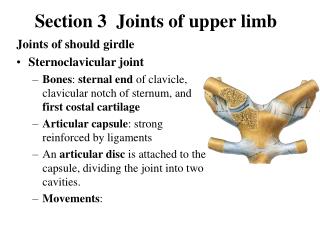
Joints of upper limb. By Dr. Eman AbdelGhany. Joints of Upper Extremity. 2. 1. 1-Sternoclavicular Synovial-saddle 2- Acromioclavicular Synovial-plane 3- Glenohumeral joint(shoulder) Synovial-ball&socket. 3. 1- Sternoclavicular joint. Articular surfaces:

Joints of upper limb
E N D
Presentation Transcript
Joints of upper limb By Dr. Eman AbdelGhany
Joints of Upper Extremity 2 1 • 1-Sternoclavicular • Synovial-saddle • 2- Acromioclavicular • Synovial-plane • 3- Glenohumeral joint(shoulder) • Synovial-ball&socket 3
1- Sternoclavicular joint • Articular surfaces: - sternal end of the clavicle. - clavicular notch of manubrium sterni. - first costal cartilage. • Ligaments: - costoclavicular ligament. - interclavicular ligament. - ant. & post. Sternoclavicular ligaments. • Blood supply: suprascapular artery & internal thoracic artery. • Nerve supply: medial supraclavicular nerve & nerve to subclavius. • NB: It is divided by articular disc into two cavities.
2- Acromioclavicular joint • Articular surfaces : - acromial end of the clavicle - lateral side of acromion process. • Ligaments: • Acromioclavicular ligament. - coracoclavicular ligament ( conoid & trapezoid parts) . • Blood supply : suprascapular & thoracoacromial arteries. • Nerve supply : suprascapular & lateral supraclavicular nerves. NB: Incompletely divided by articular disc into two compartments. Movements of both joints allow scapular rotation up to 60 degrees.
Elbow joint • Elbow Joint type: • Synovial – hinge • Articular surfaces: • Humeroulnar ( trochla of humerus+ trochlear notch of ulna ) • Humeroradial (capitulum of humerus + upper surface of head of radius) Capsule and synovial membrane are common for elbow & SRUJ Capsule is attached to humerus, ulna & unular ligament. It is not attached to radius. It includes the radial, coronoid & olecranon fossae of the humerus.
Ligaments of the Elbow 1- Ulnar Collateral Ligament medial side – connects humerus to ulna (3 bands ant. , post. And oblique bands) 2- Radial Collateral Ligament Lateral side – connects humerus to radius 3- Annular Ligament Surrounds radial head/holds it tight to ulna
Movement • Muscles Affecting the Elbow: • Elbow Flexors 1. Brachialis 2. Biceps brachii 3. Brachioradialis • Elbow Extensors 1. Triceps 2. Anconeus
Relations of elbow joint 1-Anterior: cubital fossa; Contents • Median Cubital Vein+epitrochlear lymph node • Brachial & radial + ulnar arteries • Median & radial nerves • Biceps tendon • Boundaries: Medial = Pronator teres Lateral = Brachioradialis Superior = Line between Epicodyles Roof = skin, fascia +bicipital aponeurosis Floor = supinator + brachialis muscles Frolich, Human Anatomy,UpprLimb
Relations of elbow joint ( cont.) 2- Posterior relation : triceps and anconeus muscles. 3-Medial relation : ulnar nerve & common flexor origin. 4-Lateral relation : supinator & common extensor origin. Blood supply : anastomosis around the elbow joint. Nerve supply : musculocutanious + radial + ulnar nerves.
Joints • Radioulnar joint • Radial head rotates around at proximal ulna • Distal radius rotates around distal ulna • Annular ligament maintains radial head in its joint • Joint between shafts of radius & ulna held tightly together between proximal & distal articulations by an interosseus membrane (syndesmosis) • substantial rotary motion between the bones The Elbow and Radioulnar Joints
Joints of Upper Extremity • Proximal Radioulnar joint • Synovial - pivot • Distal Radioulnar joint • Synovial – pivot • Allows pronation and supination of forearm • MiddleRadioulnar joint
Radioulnar Joint • Proximal (superior)radioulnar joint: • articulation between circumference of head of radius and radial notch of ulna . • not part of “hinge” joint • Synovial (pivot) joint • allows for forearm pronation/supination • Distal ( inferior) radioulnar joint: • articulation between head of ulna and ulnar notch of radius. • Not part of wrist joint separated from it by articular disc. • Synovial pivot joint. • allows for forearm pronation/supination • Middle radioulnar joint : fibrous syndesmosis, connect radius & ulna by interosseous membrane,
Muscles of radioulnar joints • Radioulnar pronators • Pronator teres • Pronator quadratus • Brachioradialis( begin movement ) • Radioulnar supinators • Biceps brachii(powerful supination for flexed elbow) • Supinator muscle • Brachioradialis( begin movement)
Radial styloid • Scaphoid • Lunate • Triquetral • Pisiform • Trapezium • Trapezioid • Capitate • Hamate • Metacarpal • Proximal phalanx • Middle phalanx • Distal phalanx • ulna styloid
Joints of the Upper Extremity • Radiocarpal joint(wrist): • Synovial-ellipsoid • Distal radius with proximal row of carpals(lunate & scaphoid) • Intercarpal joints • Synovial-plane • Carpal-metacarpal (2-5) • Synovial-plane • Trapezium-metacarpal 1st • Synovial-saddle • Metacarpal-phalangeal • Synovial-condyloid • Interphalangeal • Synovial-hinge
Wrist Articulations • Radiocarpal Joint • Proximal portion • Distal portion • Most surface contact found
Wrist (radiocarpal ) joint • Articulation between (lower end of radius + scaphoid & lunate ), (triquetral +articular disc)=== the ulna not share in the wrist joint . • Ligaments : 1- Ulnar collateral (styloid process of ulna to triquetral & pisiform); 2- Radial collateral ligament (styloid process of radius to scaphoid bone) 3- Ant. & post. Radiocarpal ligaments= thickened capsule. Relations: Ant. : carpal tunnel ? Post. : Extensor compartments? Lat.: Anatomical snuff box? Med.: Dorsal branch of ulnar nerve.
Extensor Compartments • Anatomic snuffbox: • EPL and EPB • Scaphoid on floor • Radial a. inside
Movement of the wrist joint • Wrist Extensors (innervated by radial n.) • Superficial • Extensor carpi radialis brevis/longus • Extensor carpi ulnaris • Extensor digitorium • brachioradialis • Deep compartment • Extensor pollicus longus/brevis • Abductor pollicus longus • Extensor indices • supinator • Secured by extensor retinaculum
Wrist flexors (median n.) • Superficial • Flexor carpi radialis • Palmaris longus • Flexor carpi ulnaris • Flexor digitorium superficialis • Pronator teres • Deep • Flexor digitorium profundus • Flexor pollicis longus • Pronator quadratus
Carpal Tunnel • Fibro-osseous structure • Floor is proximal carpal bones • Roof is transverse carpal ligament • Tunnel contains 10 structures • Median n., flexor pollicis longus tendon, 4 slips of flexor digitorium superficialis, flexor digitorium profundus • Compression results in paresthesia 2-4 fingers and decrease grip
Hand Anatomy Tendon Sheaths • Palmer Aspect
Wrist Anatomy Tendon Sheaths • Dorsal Aspect
Movement of wrist joint • Flexion: flexor carpiradialis & ulnaris + palmaris longus helped by; FDS,FDP,FPL& ABD. P.L. • Extension: extensor carpiradialis longus+ brevis & extensor carpiulnaris, helped by extensors of fingures. • Abduction: flexor carpiradialis+extensor carpiradialis longus & brevis, helped by Abd. P.L. + Ext.P.L.& brevis • Adduction: flexor & extensor carpiulnaris. • Circumduction : combination of flexion , abduction, extension & adduction. • Blood supply : palmar & dorsal carpal branches of radial and ulnar arteries & palmar arches. • Nerve supply : ant. & post. Interosseus nerves.
Joints of hand and fingures • Intercarpal: plane synovial • Carpometacarpal joint of (2-5): plane synovial • Carpometacarpal joint of thumb: synovila saddle • Intermetacarpal joints: plane synovial • Metacarpophalangeal joints: synovial condyloid • Interphalangeal joints : synovial hinge
Interphalangeal Joints of 2-5 Fingers • Hinge Joints • Motions
Ligaments of the Hand • Palmar carpometacarpal ligaments • Palmar metacarpal ligaments • Deep transverse metacarpal lig
Ligaments of the Digits • Collateral lig loose in ext, tight in flexion, obliquely • Volar plate limits hyperextension • hinged joints (uniaxial) – F/E
Stability of radius to ulna • Interosseus membrane. • Annular ligament. • Disc of inferior radioulnar joint. • Oblique cord. • Supinator & pronator quadratus muscles.
Forces transmitted from the upper limb to axial skeleton pass through: • Glenoid cavity. • Coracoid process. • Clavicle. • Costoclavicular ligament. • 1st rib & sternum. And neither through acromioclavicular joint nor through sternoclavicular joint.