Download
1 / 47
490 likes | 755 Views
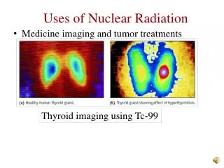
Engineering Better Medicine New Imaging Methods. D.T. Delpy Dept of Medical Physics & Bioengineering UCL. Imaging Modalities. X Ray - Conventional, Tomography Endoscopy Magnetic Resonance Imaging - MRI Single Photon Emission CT -SPECT Positron Emission Tomography - PET Ultrasound

E N D
Engineering Better MedicineNew Imaging Methods D.T. Delpy Dept of Medical Physics & Bioengineering UCL
Imaging Modalities X Ray - Conventional, Tomography Endoscopy Magnetic Resonance Imaging - MRI Single Photon Emission CT -SPECT Positron Emission Tomography - PET Ultrasound Laser Doppler Optical Coherence Tomography - OCT NIR Topography, Tomography Exogenous fluorophores MID IR Thermoacoustics Terahertz
3D X Ray CT of hand 3D X Ray CT of abdomen with soft tissue rendering
3D visualisation CT virtual colonoscopy
Images obtained when passing through the bowel (Given Imaging System)
Conventional MRI system Open access MRI system Small Extremity MRI
fMRI images of the brain showing activation changes during 40 minutes of motor learning MRI Brain image
MRI movie of the beating heart Intravascular imaging with micro-coils
MR Angiography Gated MR Image of the heart
Single headed Gamma camera PET system
Combined PET & MRI 3D image of brain Combined X ray CT and PET
Vaginal ultrasound image of early foetus 3D vaginal ultrasound image of early foetus
OCT image of the cornea and pupil OCT image of the retina
OCT of normal esophagus, colon and colon carcinoma Doppler OCT of retinal blood vessels
Absorption Spectrum of Blood ULTRAVIOLET VISIBLE NEAR INFRARED NIR Measurement Region Absorption [cm-1 M-1] HbO2 Hb Wavelength [nm]
NIR Breast Imager (Philips) Tumour Cyst Tumour 3D Breast Cup
~ ns t ~ ps t t t The MONSTIR Project But near infrared light scatters in tissue. The (temporal) Impulse Response Function of tissue to a pulse of laser light can be used to distinguish between the effects of scatter and absorption.
Arm Imaging Results (Absolute) A B A B Longitudinal MRI MRI ms ma
Optical Imaging of Luciferase tagged bacteria & tumour cells
Optical imaging of GFP fluorescence Tumour angiogenesis GFP expression monitored non invasively
Mid Infrared (900-1800 cm-1) Images of Bone and Single Osteon top: hydroxyapatite middle: Protein (amide) bottom: crystallinity
Thermoacoustic imaging of kidney with cyst Contrast depends on absorption of particular energy type
Just a few imaging topics not mentioned: X ray: spiral CT, phase imaging MRI: diffusion, perfusion, oxygenation, CSI, hyperpolarised gas blood flow PET: combined CT/PET, MRI/PET Ultrasound: power doppler, microscopy, bubble contrast (all 3D) Ultrasound/MRI: Hall effect imaging Ultrasound/Light/Microwaves: photoacoustic, thermoacoustic imaging Light: intravascular OCT, OCT spectroscopy, Raman imaging, elastic scattering spectroscopy, diffusive wave spectroscopy, polarisation imaging (structural), NIR breast imaging, flow using ICG etc etc etc
The futures bright: The futures: IMAGING
Ultrasound image and Doppler signal from umbilical artery & vein
The MONSTIR Imaging System BACK SIDE FRONT