Download
1 / 45
450 likes | 489 Views
Explore the components and classification of muscular tissue, including skeletal, cardiac, and smooth muscles. Learn about the structure and function of sarcomeres, myofilaments, and myofibrils. Study the organizational hierarchy and sliding filament hypothesis in muscle contraction.

E N D
Muscular Tissue Jun Zhou (周俊) School of Medicine, Zhejiang University 20140415
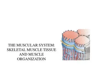
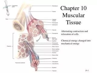
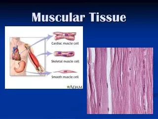
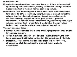
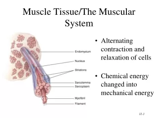
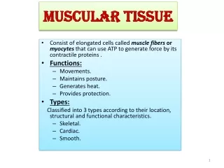
Overview 1) components: --cell: muscle fiber-myofiber • elongated thread-liked • Sarcolemma(肌膜) • Sarcoplasm(肌质) • Sarcoplasmic reticulum(肌质网): SER --extracellular G.S: CT with BV, LV and N
2) classification According to the structure and function • skeletal muscle (骨骼肌): striated, voluntary • cardiac muscle (心肌): striated, involuntary • smooth muscle (平滑肌): unstriated, involuntary
Epimysium(肌外膜): DCT • Perimysium(肌束膜) • Endomysium(肌内膜)
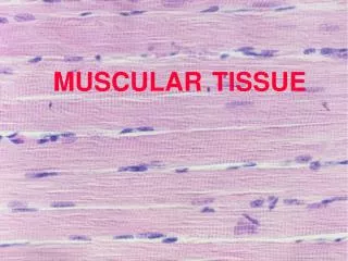
LM: • long cylindrical 10-100um in diameter, 1-40mm long • multinucleate, nuclei are ovoid, distributed under sarcolemma • filled with longitudinal parallel- arranged myofibrils
Skeletal Muscle • Cells are long multinucleate “cylinders”
Endomysium Fascicle Myofiber Perimysium Identify: Endomysium, Perimysium, myofiber and fascicle
Endomysium: connective tissue, nutrient capillaries, nerve endings and around each myofiber a basal or external lamina
Myofibril: • 1-2um in diameter • cross striation: • light band -I band • dark band- A band --A band: M-line, H-band --I band: Z-line
Definitions Isotropic (各向同性) Anisotropic (各向异性)
A band : Anisotropic (各向异性) I band: Isotropic (各向同性)
Sacromere(肌节): 1/2 I +A + 1/2 I the smallest structural and functional unit of myofibril
EM: 1.Transverse tubule (T tubule): 横小管 • definition: sarcolemma invaginate into sarcoplasm to form a transverse distributed tubular system • location: A-I junctional part • function: transfer the information into cytoplasm
2.Sarcoplasmic reticulum: 肌质网 • smooth endoplasmic reticulum • longitudinal tubule ( L tubule ) • terminal cisternae (终池) • Triad (三联体): one T-tubule +two terminal cisternae • calcium pump proteins • store and release calcium ions
3.Myofibril (肌原纤维) • thick myofilament • thin myofilament
Skeletal Muscle • Organizational Hierarchy Muscle is composed of fascicles Fascicle (smallest) is composed of myofibers Myofiber is composed of myofibrils Myofibril is composed of myofilaments
Thick myofilament • 1.5 um long, 15 nm in diameter • composed of myosin (肌球蛋白) -rob: in bundles -head: cross bridge- binding site (ATPase activity)
Thin myofilament • 1 um long, • 5 nm in diameter A. Actin (肌动蛋白)- binding site B. Tropomyosin (原肌球蛋白) C. Troponin (肌原/钙蛋白) -Tn T: bind to tropomyosin -Tn C: binds to calcium ions -Tn I: inhibit actin-myosin interaction
Myofibril Is cross banding evident at the level of the myofibril? Define a sarcomere. A sarcomere is that portion of the contractile apparatus between and attaching to two Z lines.
Myofibril Thick myofilaments consist of myosin. Myosin is ONLY found in the A Band The sarcomere is the smallest contractile unit. It consists of a precisely organized array of myofilaments.
Myofibril Thin myofilaments consist of Actin, tropomyosin and troponin. Thin myofilaments are found in BOTH the A Band and I Band
Myofibril M Line, in the center of the H Band, consists of proteins that hold myosin filaments together.
Myofibril Z Line.
Electron Micrograph of Human Skeletal Muscle Myofibril Dark granules are glycogen Micrograph Provided by Ernest Whitter
Electron Micrograph of Human Skeletal Muscle Sarcomere Micrograph Provided by Ernest Whitter
Electron Micrograph of Human Skeletal Muscle A Band Micrograph Provided by Ernest Whitter
Electron Micrograph of Human Skeletal Muscle Z Line M Line in H Band I Band Micrograph Provided by Ernest Whitter
LM • short column in shaped, 100um long,15um in diameter, with branches • 1-2 ovoid nuclei, centrally-located • striated, but no very clear • intercalated disc
EM • T tubule at Z-line level • less terminal cisternae • diad(二联体): one T-tubule + one terminal cisternae • Numerous mitochondria
LM • elongated, spindle-shaped cells, 8um in diameter, 200( 20-500)um long • rob-liked or ovoid nucleus • no striation, no myofibril
Cross Section Longitudinal Section Smooth Muscle
EM • caveola: sarcolemme invaginate into cytoplasm • dense patch (密斑): under sarcolemma dense body (密体): in sarcoplasm • intermediate filament: /composed of desmin(结蛋白) /10nm in diameter /connect between dense body
thick filament: myosin, 15 nm thin filament: actin, 5 nm-fixed on dense patch or body • contractile unit (肌丝收缩单位): several thick filament and thin filament aggregate