Download
1 / 1
10 likes | 126 Views
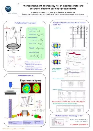
j. 0 = 0.045 m a = 0.35 m. F = 427 Vm -1. = 0,926 ± 0,002 cm -1. R. R max. Linear regression to zero kinetic energy. D. . neutral atom. F. C. h . U. e A. negative ion. 7. 8. 6. 10. 4. 5. 12. 2. 9. 1. 11. 13. 3. 14 : Laser

E N D
j 0 = 0.045 m a = 0.35 m F = 427 Vm-1 = 0,926 ± 0,002 cm-1 R Rmax Linear regression to zero kinetic energy D neutral atom F C h U eA negative ion 7 8 6 10 4 5 12 2 9 1 11 13 3 14 : Laser 15 : Column of constant F 16 : MCP 17 : Phosphor screen 18 : CCD 4 : Wien filter 7 : Deflection 11 : Focalisation quadrupole 12 : Deceleration plates 13 : Interaction zone 1 : Source and simple lens doublet (“Einzellens") 2,5,9,10 : Deflection plates 3,6,8 : Simple lenses Photodetachment microscopy to an excited state and accurate electron affinity measurements C. Blondel, C. Delsart, C. Drag, R. J. Peláez & M. Vandevraye Laboratoire Aimé-Cotton, bât. 505, CNRS, université Paris-sud, F-91405 Orsay cedex, France Photodetachment microscopy to an excited state: 31P- Photodetachment microscopy hν a Level scheme Classical parameters Highest height Maximum radius z0 = Quantum parameters : Wavelength scale Number of rings Interfrange distance Radial current density Principle: Y.N. Demkov et al., JETP Lett. 34 (1981) 403 Photodetachment microscopy: C. Blondel et al., Phys. Rev . Lett. 77 (1996) 3755 Photoionization microscopy: C. Nicole et al., Phys. Rev . Lett.88 (2002) 133001 Molecular photodetachment microscopy : C. Delsart et al., Phys. Rev . Lett.89 (2002) 183002 Experimental set-up Measured thresholds Experimental spots Deduced intervals Electron affinity 0.746 607 (10), formerly 0.746 68 (6) eV Neutral atom spectroscopy and fine structure 15 Photodetachment microscopy of Se- 14 Electron affinity : 1 629 727.4(8) m-1 formerly 1 629 780(20) m-1 or 2.020607(1) vs. 2.02067(2) eV 43rd EGAS, Fribourg, 28 June – 2 July 2011