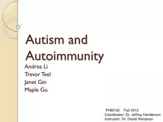
Hypersensitivity and Autoimmunity
Hypersensitivity and Autoimmunity. Hypersensitivity : Inappropriate immune reaction resulting in tissue damage Allergy : hypersensitivity reaction to an extrinsic or Intrinsic antigen The factors causing hypersensitivity called Allergen Drug Allergy

Hypersensitivity and Autoimmunity
E N D
Presentation Transcript
Hypersensitivity: Inappropriate immune reaction resulting • in tissue damage • Allergy: hypersensitivity reaction to an extrinsic or Intrinsic antigen • The factors causing hypersensitivity called AllergenDrug Allergy • (eg.Penicillin, sulphamide, asprin). • Immune system ---Protect the Immune system but hypersensitivity • Reaction---kill /damage the host cell. • Hypersensitivity does not occur in all human beings.. • Only 10% population suffer
Factors Causing Hypersensitivity • Hypersensitivity brings its symptoms either IMMEDIATE with in a minute or DELAYED , after 24-48 hrs • Hypersensitivity reaction caused by extrinsic or Intrinsic antigen • Drug--- eg.Penicillin, sulphamide, asprin • Airborne particles--- Pollen grains, house dust, spores, animal danders • Food Stuff----Shell fish, brinjal, strawbrries • Infectiousorganisms---- Bacteria, fungus, Virus, Parasite • Blood transfusion of mismatched blood
Symptoms • Diarrhoea and vomitting • Rashness • Itching • Joint pain • Fever, • Abdominal pain • Inflamation • Etc,,,,,,,,,,,,,,,,,,,,,,,,
Two types The reaction caused Hypersensitivity may be LOCAL REACTION (symptoms appear in a restricted area) Or SYSTAMATIC Reaction (symptom affect all the ORGAN system of the body) The following are common hypersensitivity reaction • Anaphylaxis • Transfusion reaction • Erythroblastsis foetails • Arthus raction • Serum sickness • Mantoux reaction • Contact dermatitis • Graves diseases
Hypersensitivity---- classified in to 2 types---- based on time reaction • IMMEDIATE Hypersensitivity • Reaction appear Immediate and disappearRAPIDLY • Inflammatory response occur FEWMINUTES • It Involves Ag-Ab interaction • Its handled by B-Cells by the production of Ab, Hence immediate hypersensitivity is Ab Mediated • It can be passivelytransferred from one host to another by the SERUM • Its INHIBITED byantihistamine Drugs
DELAYED Hypersensitivity • After 24-72 hrs • Eg. Mantoux reaction (Primarycomplex (Initial stage of Tuberculosis) reaction in children -- injection of tuberculin-- in to skin of an individual—cellmediatedResponse • This appear after 24-48 hrs • The reaction are • ERTHEMA (Redness of the Skin),,,, • INDURATION Thickiening of Tissue • Its appear SLOWLY and Last longer • As it is handled by T cells, It is a CELLMEDIATED Immunity • It can not be transferred from one host to another by the SERUM • It is suppressed by CORTICO STERORIDS
IMMEDIATE hypersensitivity • Type I anaphylactic or immediate • Type II Ab Dependent toxic • Type III Complex Mediated • Type V Stimulatory • DELAYED Hypersensitivity • Type IV Cell mediated
Type I - immediate anaphylactic • The Term anaphylaxis was coined by Richet in 1902 and it means “WithoutProtection” • Theterm anaphylaxis comes from Greek word. Ana=Against (Without), Phylaxis =Protection • Type I hypersensitivity is an allergic reaction provoked by re-exposure to a specific antigen. • Exposure may be by ingestion, inhalation, injection, or direct contact. • The reaction is mediated by IgEantibodies and produced by the immediate release of histamine,tryptase, arachidonate and derivatives by basophils and mast cells..
This causes an inflammatoryresponse leading to an immediate (within seconds to minutes) reaction. • The reaction may be either local or systemic. Symptoms vary from mild irritation to sudden death from anaphylactic shock. • Treatment usually involves epinephrine,antihistamines, and corticosteroids Some examples: • Allergic asthma • Allergic conjunctivitis • hay fever
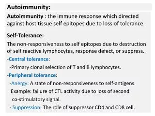
Mechanism of type I hypersensitivity pollens house dust mite animal dander foods (eg peanut) wasp / bee venom Extrinsic allergen IgE Th2 response IL-4 / IL-13 mast cells Priming sensitization elicitation
Factors causing Aanaphylaxis • Drugs-penicillin • Venom from insects (Bees) • Food • Food additives • Egg • Shell fish • Pollen grain • Temperature- Heat or Chillness
Treatment Symptoms • Avoiding contact with Allergen • Drugs • Epinephrine • Isoprenaline • Sodium chromoglycate • Itching • Reddish patches in skin • Swelling of lips • Swelling of tongue, throat, Conjunctiva • Running nose • Wheezing, cough • Low bp • Abdominal pain • Vomiting , Headache • Cardiac arrest -Death
Type II - antibody-dependentCytotoxic Hypersensitivity • In type II hypersensitivity, the antibodies produced by the immune response bind to antigens on the patient's own cell surfaces. • The antigens recognized in this way may either be intrinsic("self" antigen, innately part of the patient's cells) or extrinsic (absorbed onto the cells during exposure to some foreign antigen, possibly as part of infection with a pathogen
IgG and IgMantibodiesbind to these antigens to form complexes that activate the classicalpathway of complementactivation for eliminatingcellspresenting foreign antigens (which are usually, but not in this case, pathogens). • As a result mediators of acute inflammation are generated at the site and membraneattackcomplexes cause cell lysis and death. The reaction takes hours to a day.
Mechanism of CYTOTOXIC REACTION • In cytotoxic reaction the CELL DAMAGE occur in any one of the following methods • Phagocytosis • Lysis (Ab binds to the Ag and stimulate the complement ) • Killing (The Ab coated cells may be attack by cytotoxic killer cells (which carry C3b and Fc portion of IgG)
Type II (antibody mediated) hypersensitivity Antibody to tissue bound or cellular antigen:
Examples • Agglutination and lysis of blood cells due to mismatch • Autoimmune haemolytic anaemia • Immune thrombocytopenia • Transfusion reactions • Hashimoto's thyroiditis • Graves' disease • Myasthenia gravis • Hemolytic disease of the newborn
Types of Cytotoxic Hypersensitivity • Cytotoxic Hypersensitivity classified in to 2 Types • ISOIMMUNEREACTION (Reactionbring about Ag-Ab of TWO INDIVIUDALS belonging to the same Species. Eg.Blood group Ag and Leukocyte (WBC) Ag. • AUTOIMMUNEREACTION (Reactionbring about by the interaction b/w Ag-Ab of the same individual
ISOIMMUNEREACTION • ISOIMMUNEREACTION (Reactionbring about Ag-Ab of TWO INDIVIUDALS belonging to the same Species. Eg.Blood group Ag and Leukocyte (WBC) Ag. • Transfusion Reaction (ABO Grouping) • Erythroblastosis fostalis • Rh+ baby born to Rh-mother first time fine. 2nd time can have abs to Rh from 1st pregnancy. • Ab crosses placenta and baby kills its own rbcs. • Treat mother with ab to Rh antigen right after birth and mother never makes its own immune response. • Transpalnt rejection reaction
AUTOIMMUNEREACTION (Reactionbring about by the interaction b/w Ag-Ab of the same individual • Autoimmune reaction due to interaction of Auto Ag and Ab • Auto immune Hemolytic anemia • Auto immune thrombocytopaenic purpura • Autoimmune thyroiditis • Mysthenia gravis
Rhesus disease of the newborn – a type II hypersensitivity disease
Myasthenia gravis • It is a long term neuromuscular disease that leads to varying degrees of muscle weakness • The most commonly affected muscles are those of the eyes, face • It can result in double vision, drooping eyelids, trouble talking, and trouble walking • Myasthenia gravis is an autoimmune disease which results from antibodies that block acetylcholine receptors at the junction between the nerve and muscle • This prevents nerve impulses from triggering muscle contractions
Myasthenia gravisthe mechanism • Myasthenia gravis is an autoimmune disease which results from antibodies that block acetylcholine receptors at the junction between the nerve and muscle
Myasthenia gravis SYMPTOMS • Symptoms include weakness in the arm and leg muscles, double vision and difficulties with speech and chewing. • Muscular: muscle weakness or weakness of the arms and legs • Facial: drooping of upper eyelid or muscle weakness • Also common: difficulty swallowing, double vision, fatigue, shortness of breath, weak eye muscles, or impaired voice
Grave’s disease Thyrotrophic stimulate hormone (TSH)
Graves' disease SYMPTOMS • Symptoms include anxiety, hand tremor, heat sensitivity, weight loss, puffy eyes and enlarged thyroid. • Tremor: in the hands • Whole body: excess sweating, excessive hunger, fatigue, heat intolerance, high blood pressure, or sweating • Heart: fast heart rate, irregular heart rate, or sensation of an abnormal heartbeat • Eyes: abnormal projection of eyes, puffy eyes, or watery eyes • Anxiety or nervousness • Also common: absence of menstruation, diarrhea, enlarged thyroid, graves' ophthalmopathy, hair loss, insomnia, irritability, overactive thyroid, or weight loss
Type III - immune complex In type III hypersensitivity: • Soluble immune complexes (Ag-Ab) (aggregations of antigens and IgG and IgM antibodies) form in the blood and are deposited in various tissues (typically the skin, kidney, lungs and joints) • This may trigger an immune response according to the classical pathway of complement activation. • The reaction takes hours to days to develop
Mechanism • The mechanism includes T lymphocytes and monocytes and/or macrophages. • Cytotoxic T cells (Tc) cause direct damage whereas helper T (TH1) cells secrete cytokines which activate cytotoxic T cells, recruit and activate monocytes and macrophages, which cause the bulk of the damage • The delayed hypersensitivity lesions mainly contain monocytes and a few T cells.
Type III (immune complex) mediated hypersensitivity
The classical example of this hypersensitivity is tuberculin (Montoux) reaction • Reaction peaks 48 hours after the injection of antigen (Old tuberculin). The lesion is characterized by induration and erythema
Examples: • Rheumatoid arthritis • (Arthritis of joints involves inflammation of the synovial membrane. Joints become swollen, tender and warm, and stiffness limits their movement. • Most commonly involved are the small joints of the hands, feet and cervical spine • They occur in 30% of people • Serum sickness • Symptoms of malaria • Systemic lupus erythematosus • Arthus reaction
Early and late joint changes in rheumatoid arthritis
Rheumatoid arthritis SYMPTOMS • Pain areas: in the back, joints, or muscles • Joints: stiffness, swelling, tenderness, or weakness • Whole body: anaemia, fatigue, or malaise • Skin: lumps or redness • Hand: bump on the finger or swelling
Systemic lupus erythematosus • Is an autoimmune disease in which the body’s immune system mistakenly attacks • healthy tissue in many parts of the body • Female sex hormones, sunlight, smoking, vitamin D deficiency, and certain • infections, are also believed to increase the risk • The mechanism involves an immune response by autoantibodies (auto antibodies • that bind to contents of the cell nucleus) against a person's own tissues
Systemic lupus erythematosusSymptoms • Pain areas: in the eyes, joints, or muscles • Pain types: can be sharp (Prickly) in the chest • Whole body: anaemia, fatigue, fever • Mouth: dryness or ulcers • Skin: rashes on the face or red rashes • Hair: hair loss or loss of scalp hair • Also common: arthritis, blood in urine, face rash, headache, joint stiffness, lupus headache, mental confusion, sensitivity to light, swelling, swollen (puffy) lymph nodes, water retention, or weight loss
Type IV hypersensitivity • Delayed reaction • 36 to 48 hours • Characterized by induration and erythema • Also known as cell mediated (T cells) hypersensitivity
Type IV hypersensitivity “Delayed-type” -- slow onset ~day(s) (if sensitized) TH1-cell mediated Sensitization phase-- -- TH1 expansion Effector stage -- TH1 & macrophage activation -- inflammation Latex type IV hypersensitivity Hypersensitivities
Type IV hypersensitivities, con’t • Contact dermatis reactions • Often involves hapten production • e.g. to: hair sprays, plant toxins TH1 Hypersensitivities