Download
1 / 26
490 likes | 817 Views
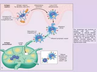
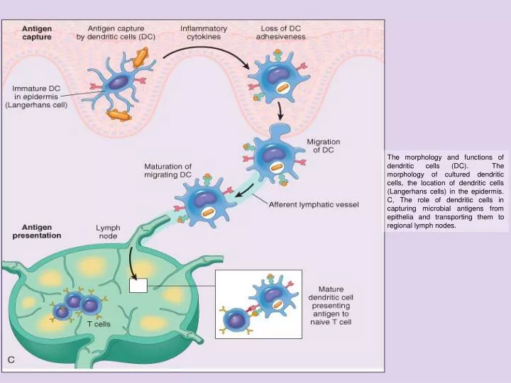
The morphology and functions of dendritic cells (DC). The morphology of cultured dendritic cells, the location of dendritic cells (Langerhans cells) in the epidermis. C, The role of dendritic cells in capturing microbial antigens from epithelia and transporting them to regional lymph nodes.

E N D
The morphology and functions of dendritic cells (DC). The morphology of cultured dendritic cells, the location of dendritic cells (Langerhans cells) in the epidermis. C, The role of dendritic cells in capturing microbial antigens from epithelia and transporting them to regional lymph nodes.
Scheme of lymphocyte development and sites of block in primary immunodeficiency diseases. The affected genes are indicated in parentheses for some of the disorders. ADA, adenosine deaminase; CD40L, CD40 ligand; SCID, severe combined immunodeficiency.
Pathogenesis of HIV-1 infection. Initially, HIV-1 infects T cells and macrophages directly or is carried to these cells by Langerhans cells. Viral replication in the regional lymph nodes leads to viremia and widespread seeding of lymphoid tissue. The viremia is controlled by the host immune response (not shown), and the patient then enters a phase of clinical latency. During this phase, viral replication in both T cells and macrophages continues unabated, but there is some immune containment of virus (not illustrated). There continues a gradual erosion of CD4+ cells by productive infection (or other mechanisms, not shown). Ultimately, CD4+ cell numbers decline, and the patient develops clinical symptoms of full-blown AIDS. Macrophages are also parasitized by the virus early; they are not lysed by HIV-1, and they may transport the virus to tissues, particularly the brain.
Schematic illustration of an HIV-1 virion. The viral particle is covered by a lipid bilayer that is derived from the host cell. Mechanisms of CD4 cell loss in HIV infection.
Pathogenesis of autoimmunity. Autoimmunity results from multiple factors, including susceptibility genes that may interfere with self-tolerance and environmental triggers (inflammation, other inflammatory stimuli) that promote lymphocyte entry into tissues, activation of lymphocytes, and tissue injury.
Role of infections in autoimmunity.Infections may promote activation of self-reactive lymphocytes by inducing the expression of costimulators (A), or microbial antigens may mimic self-antigens and activate self-reactive lymphocytes as a cross-reaction (B).
Role of infections in autoimmunity. Infections may promote activation of self-reactive lymphocytes by inducing the expression of costimulators (A), or microbial antigens may mimic self-antigens and activate self-reactive lymphocytes as a cross-reaction (B).
Reazioni immunopatogene (malattie da ipersensibilità) Reazioni immunopatogene (malattie da ipersensibilità) Ipersensibilità (o allergia): qualsiasi reazione immunitaria capace di produrre un danno nei soggetti predisposti immunopatogene (malattie da ipersensibilità). non significa risposta immunitaria esagerata ma piuttosto una risposta immunitaria che di per sé è capace di danneggiare il tessuto dell'ospite e di provocare malattie da ipersensibilità In base al loro meccanismo patogenetico si distinguono 4 tipi principali (in realtà i processi immunitari delle reazioni d'ipersensibilità sono in parte sovrapposti): di tipo I – Ipersensibilità immediata o anafilassi di tipo II – Ipersensibilità mediata da anticorpi citotossici reazioni di tipo immediato di tipo III – Ipersensibilità mediata da immunocomplessi di tipo IV – ipersensibilità di tipo ritardato o cellulo-mediata reaz. di tipo ritardato, che appare dopo ore o giorni
Pathogenesis of immediate (type I) hypersensitivity reaction. The late-phase reaction is dominated by leukocyte infiltration and tissue injury. TH2, T-helper type 2 CD4 cells.
Schematic illustration of the three major mechanisms of antibody-mediated injury.A, Opsonization of cells by antibodies and complement components and ingestion by phagocytes. B, Inflammation induced by antibody binding to Fc receptors of leukocytes and by complement breakdown products. C, Antireceptor antibodies disturb the normal function of receptors. In these examples, antibodies against the thyroid stimulating hormone (TSH) receptor activate thyroid cells in Graves disease, and acetylcholine (ACh) receptor antibodies impair neuromuscular transmission in myasthenia gravis.
Schematic illustration of the three major mechanisms of antibody-mediated injury. A, Opsonization of cells by antibodies and complement components and ingestion by phagocytes. B, Inflammation induced by antibody binding to Fc receptors of leukocytes and by complement breakdown products. C, Antireceptor antibodies disturb the normal function of receptors. In these examples, antibodies against the thyroid stimulating hormone (TSH) receptor activate thyroid cells in Graves disease, and acetylcholine (ACh) receptor antibodies impair neuromuscular transmission in myasthenia gravis.
Schematic illustration of the three sequential phases in the induction of systemic immune complex-mediated disease(type III hypersensitivity).
Pathogenesis of immune complex-mediated tissue injury. The morphologic consequences are depicted as boxed areas.
Advanced systemic sclerosis. The extensive subcutaneous fibrosis has virtually immobilized the fingers, creating a clawlike flexion deformity. Loss of blood supply has led to cutaneous ulcerations.
Mechanisms of T cell-mediated (type IV) hypersensitivity reactions. A, In delayed type hypersensitivity reactions, CD4+ T cells (and sometimes CD8+ cells) respond to tissue antigens by secreting cytokines that stimulate inflammation and activate phagocytes, leading to tissue injury. B, In some diseases, CD8+ cytolytic T lymphocytes (CTLs) directly kill tissue cells. APC, antigen-presenting cell.
Mechanisms of T cell-mediated (type IV) hypersensitivity reactions. A, In delayed type hypersensitivity reactions, CD4+ T cells (and sometimes CD8+ cells) respond to tissue antigens by secreting cytokines that stimulate inflammation and activate phagocytes, leading to tissue injury. B, In some diseases, CD8+ cytolytic T lymphocytes (CTLs) directly kill tissue cells. APC, antigen-presenting cell.
Schematic illustration of the events that give rise to the formation of granulomas in cell-mediated (type IV) hypersensitivity reactions. Note the role played by T cell-derived cytokines.
trapianto: trasferimento di cellule, di tessuti e di organi da un individuo donatore a un ricevente o ospite; gli antigeni responsabili del rigetto dei trapianti sono quelli codificati dai geni MHC HLA (Human Leukocyte Antigens) = sistema identificato nell'uomo sul cromosoma 6 che comprende i geni che codificano per gli antigeni di istocompatibilità (→ Complesso maggiore di Istocompatibilità, MHC). Locus (pl. loci): posizione di ciascun gene all'interno di un sistema (porzione di cromosoma);
Diversi tipi di rigetto dei trapianti Tipo di rigetto Tempo Meccanismo primario di rigettoiperacuto 5–30 minuti ipersensibilità di tipo II acuto 4-30 giorni immunità cellulo-mediata ipersensibilità di tipo IV cronico > di 3 mesi probabili reazioni anticorpo-mediate ipersensibilità di tipo III
Fattori che influenzano il trapianto Favorevoli Sfavorevoli Buon appaiamento dei sistemi HLA + BO Infezioni Tolleranza indotta da precedenti trasfsioni Tossicità ai farmaci Uso di farmaci immunosoppressori Malattie ricorrenti
Schematic illustration of the mechanisms involved in central and peripheral tolerance. The principal mechanisms of tolerance in CD4+ T cells are shown. APC, antigen-presenting cell.
Schematic representation of the events that lead to the destruction of histoincompatible grafts. In the direct pathway, donor class I and class II antigens on antigen-presenting cells in the graft (along with B7 molecules, not shown) are recognized by CD8+ cytotoxic T cells and CD4+ helper T cells, respectively, of the host. CD4+ cells proliferate and produce cytokines that induce tissue damage by a local delayed hypersensitivity reaction and stimulate B cells and CD8+ T cells. CD8+ T cells responding to graft antigens differentiate into cytotoxic T lymphocytes that kill graft cells. In the indirect pathway, graft antigens are displayed by host APCs and activate CD4+ T cells, which damage the graft by a local delayed hypersensitivity reaction. The example shown is of a kidney allograft.