Download
1 / 47
480 likes | 652 Views
Learn about the immune system's role in distinguishing between self & non-self, and the mechanisms of defense against pathogens and transformed structures. Explore antigen processing, activation of innate system, and MHC functions.

E N D
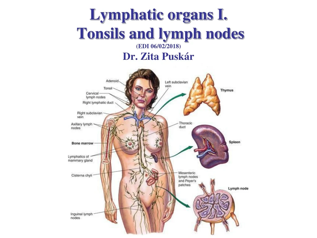
Lymphatic organs I. Tonsils and lymph nodes(EDI 06/02/2018)Dr. Zita Puskár
The immune system Function: distinguishing between self or non-self, dangerous or non-dangerous and responding to those with tolerance or elimination.4 Immunohomeostasis - maintenance of the genomic permanency • Defense against the pathogens • Elimination of the transformed self structures Antigen: every structure (cells, molecules, microbes) that the immune system recognizes and responds to it
Innate and adaptive immune system Innate Cells → cellular immune response Monocytes - macrophages Granulocytes Dendritic cells Mast cells Soluble molecules → humoral immune response Complement proteins (glycoproteins, enzymes, receptors) Adaptive Cells → cellular immune response B lymphocytes T lymphocytes Soluble molecules → humoral immune response Antibodies
Activation of the innate system Recognized structures: Pathogen Associated Molecular Pattern (PAMP) Receptors: Pattern Recognizing Receptors (PRR). They are not clonal. They are the same on different cell types. Fc receptors, binding antigen-antibody complexes. PPR (Pattern recognition Receptors) (only examples) Mannose receptor Scavenger receptor CD 14 Toll-like receptors
Antigen processing Phagocytosis, antigen presentation, cell activation – „professional” antigen presenting cells (APCs): macrophage, dendritic cell • Phagocytosis, antigen presentation, cell activation oxygen burst (bacterial infections) – neutrophil granulocyte (microphage) • Elimination of helminthic worms and protozoa, (MBP toxicity, ECP neurotoxicity), allergic reaction with inflammation – eosinophil granulocyte • Allergic reaction, defence against parasites – mast cell, basophil granulocyte • Elimination of viral infected and tumour cells – Natural Killer Cell (NK)
Interaction between the innate and adaptive immune systems Immune response Patogens, Antigens Innateimmunesystem Innate complementactivation mastcell granulocyte Humoral Cellular Adaptive plasma cell lymphokines Innate complement Activated Mf lysis lysis phagocytosis
Major Histocompatibility Complex:MHC-I and -II Function: Determination the immunological „self”, helping the formation of T-cell repertoire and the recognition of proteins by T-cells (membrane glycoproteins – peptidereceptors) Peptides that join the MHC I consist of 8-10 amino acids. Cells that express MHC-I–allnucleated cell Peptides that join the MHC II contain 13-23 amino acid. Cells that express MHC-II –B cell, macrophage, dendritic cell (APC), thymic epithel Human MHC-HLA: Human Leucocyte Antigen complex
Antigen presentation Antigens originated from lipids join to CD1, peptide antigens join to MHC-I or MHC-II. Endogenous proteins (the host cell synthesizes foreign proteins with tumour cell like characteristics in viral or bacterial infected cells). Antigen processing: proteasome (enzymes) → ER (peptide fragments join to MHC-I→ Golgi → vesicular transport → appearance on the cell surface Exogenous proteins (are taken up by APCs by fagocytosis, pinocytosis or receptor mediated endocytosis) Antigen processing: endo- and lysosome system → protected MHC II - formed in the ER join to vesicular system → protein binds to MHC-II → appearance on the cell surface
Activation of NK cells NK cells express „Killer Activating Receptor (KAR) „ and „Killer Inhibitory Receptor” (KIR). Binding „self” MHC-I molecules to the KIRs inhibitsthe KARs. NK cell does not destroy self. Virus infected and tumour cells decrease the MHC-I expression to hide themselves from cytotoxic T-cells. Therefore there is no enough MHC-I to activate the KIR so the NK cells attack.
Nomenclature CD (Clusters of Differentiation) – nomenclature of the cell surface molecules that determine the type of the cell, the stage of the differentiation or activation. Cell line markers (eg. Hemopoietic stem cell CD34+, T-helper cell - CD4+, T-citotoxic-CD8+ • Maturation markers (Tymocytes in the thymus CD1+, mature T lymphocyte does not express this) • Activation markers (molecules appearing after stimulation pl. CD25) Cytokines: small peptides or glycoproteins that regulate the cell functions in the immune system through receptor mediated pathways. Lymphokines-cytokines produced by lymphoid cells.
Antibodies – Immunoglobulins (Ig) IgM: naive B cell antigen binding receptor IgD : naive B cell antigen binding receptor IgG: „switched” B cell antigen binding receptor (opsonisation, complement activation, maternal immunity) IgA: „switched” B cell antigen binding receptor (mucosa immunity) IgE: „switched” B cell antigen binding receptor (immediate hypersensitivity) monomer dimer pentamer trimer
Diversity of the antigen binding receptors Genetic mechanism that results in diversity and specificity is the somatic recombination of genes that code the antigen binding site of the immunoglobulins and T-cell receptors.
B-cell receptor (BCR) and B-cell activation BCR: membrane bound immunoglobulin molecule with signal transduction chains. BCR reacts with soluble or corpuscular antigens. B-lymphocyte activation: antigen binding, interaction with macrophages and lymphokines from T helper cells result in division and differentiation of B cells into plasma cells and memory B cells.
T-cell receptor (TCR) and T-cell activation TCRs: coded by genes belonging to the Ig super-family. They recognize only linear sequence of peptide fragments that join to MHC molecules!!! TCR-MHC-antigen binding is not enough for T-cell activation. Co-stimulatory molecules are necessary!!!
T-cell classes Production of cytokines (lymphokines) – activation of macrophages, regulation of inflammatory and cytotoxic processes, defense against intracellular pathogens, cellular immune response MHC-II-exogene antigen Th1 Production of lymphokines – activation of B cells, differentiation into plasma cells, defense against extracellular pathogens, humoral immune response Th2 CD4+ Treg Production of lympholines – regulation of immune response Th: helper T-cell Treg: regulatory T-cell Tcit : cytotoxic T-cell Destroys virus or intracellular pathogen infected or tumour cells (perforin, granular enzymes.) CD8+ Tcyt MHC-I-endogeneous antigen
Organization levels of the immune system Cells: cells of the innate and adaptive immune system Tissues: blood and lymphatic tissues Organs: lymphatic organs
Organization of lymphatic organs Primary (central) lymphatic organs Bone marrow: formation of T and B lymphocyte and maturation of B-cells Thymus (thymus gland): maturation ofT-cells Secondary (peripheral) lymphatic organs: „meeting with the antigens” → activation of B and T cells Mucosa Associated Lymphatic Tissue (MALT:antigen in the mucosa) Gastriontestinal tract (digestive tract) GALTTonsils Peyer’s patches in the wall of the intestine Appendix vermiformis Nasal (NALT), Bronchoalveolar (BALT) and urogenital system associated lymphatic tissue Skin Associated lymphatic Tissue (SALT), antigen in the skin) Lymph and lymph node (antigen in tissues) Spleen (antigen in the blood)
Different forms of lymphatic tissue Diffuse lymphoid elements (lymphocytes) Solitary lymph nodule or follicle Epithelium related lymphatic tissue : tonsils Aggregated lymph nodules (pl. Payer’s plaque) Capsulated lymphatic organs spleen Lymph node thymus
General features of peripheral lymphatic organs Stroma: reticular connective tissue that consists of reticular fibers forming 3D networks and reticular cells. Immune cells and accessory cells are densely packed within the reticular connective tissue (lymphoreticular tissue) Cells: lymphoid cells: B and T lymphocytes, plasma cells, NK cells, accessory cells: macrophages, dendritic cells: follicular dendritic cells (FDC): bind native antigens and help the differentiation of B-cells in the germinal center of the lymphatic nodule (B- dependent area) dendritic cells (interdigitating dendritic cell) present MHC-II joined antigens to the a Th cells in the T- dependent area of lymphatic organs, (eg. Langerhans cells of the skin, interstitial dendritic cells in the connective tissue) Vessels: blood vessels (with high endothelial venules, HEV) lymph capillaries Special formations: lymphatic nodules (follicles)
Dendriticcell (DC) DC-s originate from the bone marrow. These cells have long processes. The nucleus is irregular, segmented and the cellular organelles surround it.
Highendothelialvenule (HEV) Postcapillary venules have unusual endothelial cells that are high cuboidal and protrude into the lumen of the vessel. The nucleus is large, spherical and lightly stained. The surface glycoproteins and integrins of the apical part of the endothels facilitate fast diapedesis of lymphocytes out of the blood. (15-20 000 cell/second)
Primer és secunderfollicles (lymphaticnodules) Cap Reticular cell FDC Light zone Germinal center Primer (d=50-100 µm) Germinal center reaction Dark zone Primer: reticulum cells, resting naive B-lymphocytes (antigen free environment, e.g. intrauterine life) Secunder: cap (like the primer follicle). Germinal centre (centrum germinativum): dark zone - B lymphoblasts (centroblasts), dividing forms light zone-smaller centrocytes , follicular dendritic cells, macrophages s v Secunder (d=200-400 µm)
Germinalcenter-reaction: B-cellactivation centrocyta centroblast tingible body macrophage
T- and B-dependent areas lymph node spleen tonsil Naive B-cells, macrophages, follicular dendritic cells are the main cell types in the follicles, while naive T cells, dendritic cells, macrophages are located in the interfollicular areas.
MALT: Tonsils Pharyngeal tonsil Waldeyer ring Tubal tonsil Palatine tonsil Lingual tonsil
K General features of tonsils MALT – Mucosa Associated Lymphatic Tissue : • Lymphoepithelial tissue: the epithelium is infiltrated with lymphatic cells • Lymphoreticular connective tissue with lymphatic nodules. The follicles are B-dependent areas. Among the follicles, T-dependent areas with blood vessels (HEV), and lymph capillaries. crypts (fossula, lacuna): epithelial invaginations (increasing the surface for immune response) K K lymphoepithel K K
Histology of lingual tonsil • Epithelium: stratified squamous non-keratinizing epithelium infiltrated with lymphoid cells. • Crypts are not too deep • Lymphoreticular connective tissue with follicles • Mucous lingual glands that release their saliva into the crypts • Skeletal muscle • Adipose tissue
Palatine tonsil Palatopharingeal arch tonsillar fossa Palatoglossal arch
Histology of palatine tonsil hemicapsule • Epithelium: stratified squamous non-keratinizing epithelium infiltrated with lymphoid cells. • Crypts are deep and branched (during bacterial infection (strepto- or staphylococcus) the crypts are filled with neutrophils → pus • Lymphoreticular connective tissue with follicles • Hemicapsule: a band of dense connective tissue acting as a capsule separating the lymphoid tissue from the subjacent structure. Function: barrier against to spread infections (area for tonsillectomy)
Tonsillitis, tonsillectomy physiological inflammed tonsillectomy follicular tonsillitis
Pharyngeal tonsil In the pharyngeal fornix
Histology of pharyngeal tonsil Structure: 6-8 sagittal folds and deep pits among the folds Epithelium: ciliated pseudostratified columnar epithelium (with islands of stratified squamous non-keratinizing epithelium on the top of the folds and in the pits) infiltrated with lymphoid cells.
MALT: Peyer’s patches ileum
Lymph and lymphatic vessels Lymph: interstitial (extracellular) fluid with some red blood cells and lymphoid cells. Lymphcapillary: thin endothelial cells formed tubes covered by basal lamina and connective tissue fibers. It appears almost every tissue type with few exceptions (nervous system). It contains valves that determine the direction of the lymph flow. Vasa lymphatica (larger lymph vessel, intima: endothel, elastic fiber, media: circular smooth muscle layers, adventitia collagen fibers) Lymphatic ducts : the largest lymph vessels (intima: endothel, elastic fibers, media: circular smooth muscle cells, adventitia: spirally organized smooth muscle cells mixed with connective tissue fibers valves
Pressure conditions in the lymphatic circulation P=P(cap)hydr – P(int)hydr + P(int)ko - P(cap)ko P highest P lowest Lymphokinetic motion and pressure gradient Capillary →extracellular fluid→lymph capillary→ vasa lymphatica→ lymphatic duct →large vein
Lymph and blood circulation jugular vein subclavial vein Lymph capillary → vasa lymphatica → lymphatic duct → large veins
Lymph node Capsulated lymphatic organ embedded into adipose tissue. cortex Stroma: reticular connective tissue Structure: (Cell rich) parenchyma lymph sinusoids Cortex: dense parenchyma with follicles and less sinusoids Paracortex: dense parenchyma less sinuses, no follicles Medulla: lymph sinusoids, with parenchyma (medullary cords) extending from the cortex paracortex trabecule capsule medulla
reticular cell Reticular fibers – silver impregnation
medulla cortex follicle medullary cord medullary sinus trabecule
Cortex and paracortex subcapsular sinus cortex paracortex Cortex: follicles (B dependent area) with follicular dendritic cells and follicular macrophages, interfollicular zone (T dependent area as the paracortex), Paracortex: T lymphocytes , dendritic cells, macrophages, HEV B T
Medulla medullary cord Medullary cord: B-cells, few T-cells, plasma cells Medullary sinus: flat reticular cells (sinusendothel) covered vessels in which lymph and cells are located (lymphocytes, plasma cells, macrophages)
Lymphatic circulation Lymph route vasa afferentia ↓ marginal sinus (subcapsular) ↓ trabecular sinus ↓ medullary sinus ↓ vasa efferentia Afferent lymph vessel (vasa afferentia, VA) marginal sinus capillary postcapillary venule (with HEV) VA marginal sinus medullary sinus VA trabecular sinus Gate (hilus) trabecular sinus Efferent lymph vessel vasa efferentia Blood circulation: (enter through the hilus) artery → capillary → postcapillary venule (HEV) → hilar vein
High Endothelial Venule HEV - high endothelial venule which are located mainly in the paracortex. The cuboidal or low columnar endothelial cells with large lightly stained nucleus. Circulating lymphocytes leave the blood circulation (homing, 15-20 thousand cells/second!)
Function of the lymph node afferent lymp vessel antigen Bp/Bm FDC cell carried antigen pl.Langerhans s. efferent lymph vessel B HEV Th T Ta/Tm
Regional lymph v. jugularis R L Right lymphatic duct Cervical (neck) Supra- and infraclavicular Thoracic duct Axillary (upper limb) Paraaortic lymph nodes Pelvic peripheral lymph (unfiltrated) primary lymph nodes Inguinal (lower limb) tertiary nodes lymphatic ducts secondary nodes central lymph (filtrated, cell-rich)