Understanding Departmental Measurement Criteria for Carotid Artery Stenosis Assessment
110 likes | 249 Views
This document outlines the essential departmental measurement criteria for assessing carotid artery stenosis using the ASUM guidelines. It emphasizes the importance of consistent cursor placement to obtain clear spectral windows, particularly in cases of less than 50% stenosis. Detailed calculations using NASCET and ECST criteria are discussed, highlighting the relationship between residual lumen and stenosis measurement. The distinctions between these methods are explored to underscore their clinical relevance, particularly in determining the risk associated with treatment plans.

Understanding Departmental Measurement Criteria for Carotid Artery Stenosis Assessment
E N D
Presentation Transcript
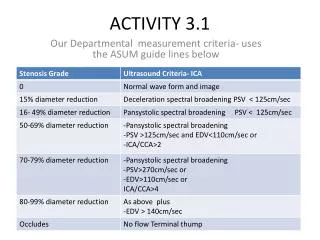
ACTIVITY 3.1 Our Departmental measurement criteria- uses the ASUM guide lines below
However there is emphasis on the need to be consistent in cases of <50% stenosis, by accurately placing the curser in mid stream to obtain a good clear spectral window-because spectral broadening can occur with increased transducer pressure and when the curser or sample volume is placed near the vessel wall as shown below.
Curser in mid stream with increased transducer pressure causing spectral broadening
Curser placed near the vessel wall again shows spectral broadening
NASCET CRITERIA Minimum residual lumen at the point of maximum stenosis referenced to the diameter of distal lumen for the internal carotid artery at the first point at which the arterial walls become parallel. The image below show plague of the ICA bulb, the residual lumen was measured as well as the normal ICA distal to the bulb.
Calculation • A= 4.7 B=5.6 therefore 1-4.7 5.0x100=0.6% Minimal stenosis- there will be no significant increase in peak systolic velocity until >50%
NASCET is more straight forward indicator of the degree to which the ICA is narrowed by the stenosis, in that it compares the stenosis to the distal lumen of the artery and is reproducible and reliably measured from the angiograms . Uses the diameter of the disease free point in ICA as the denominator The ECST method gives a better indication of plague burden at the sight of the stenosis but uses the artery diameter as the denominator at the same point
THE ECST CRITERIA • Uses a different method
I think that the ECST criteria is no longer popular because it produces higher values for modest and significant benefit being between70-99% equivalent to NASCET of 50-69%. A ECST 50% stenosis within the carotid bulb may yield 0% using the NASCET method, that is very risky to the patient ‘s treatment plan.`