Download
1 / 43
430 likes | 541 Views
Understand the physiology of blood group antigens, the significance of matching blood types in transfusions, and the implications of ABO and Rh systems in healthcare. Learn about the common antigens and antibodies present in various blood groups.

E N D
BLOOD GROUPS AND THEIR CLINICAL SIGNIFICANCE HEM_PHY Lecture 5
Why is blood needed? Blood is needed for • Accidents esp. MVA cases • Transfusions in surgery • Treatments : Leukemia, cancer, thalassemia, haemophilia, complications during delivery, etc.
Desired Learning Outcomes On completion of this topic, you should be able to • Explain the importancetypes of blood groups and discuss its clinical importance. • Describe the physiology of mismatched blood transfusion reactions • Explain the process of cross matching – major and minor
RBC membranes have glycoprotein antigens on their external surfaces. Human erythrocytes has >300 antigenic determinants These antigens are: unique to the individual promoters of agglutination and are referred to as agglutinogens recognized as ‘foreign’/non-self if transfused into another individual Presence or absence of these antigens is used to classify blood groups Human Blood Groups
Markers on RBC Agglutinogens = antigens that promote agglutination A B O – 2 glycoprotein antigens, A and B Rh Duffy; Kidd; Kell; Lewis NOTE: Agglutinogens of these systems are usually very poor antigens – they do not produce antibodies against them and reactions are relatively mild.
i. The ABO System • Discovered in 1901 by Dr. Karl Landsteiner • 4 main phenotypes (A, B, AB, O) • ABO gene located on long arm of chromosome 9. • Antigens/agglutinogens Type A and Type B on the surface of the RBCs are the cause of blood transfusion reactions [BTR] • Thus, the presence or absence of these Ag’s is the essential basis that blood is grouped for the purpose of transfusion.
Landsteiner’s Law • Only applicable to the ABO system • 2 laws : • 1st Law: When an Agglutinogen is present on the membrane of RBC, the corresponding agglutinin must be absent in the plasma of the person. Eg: RBC contains A –Ag; plasma must not contain α agglutinin; otherwise there will be death! • 2nd Law: WheN the RBCs in an individual is devoid of agglutinogen, plasma shall contain the corresponding agglutinin. Eg: RBCs contain no B or A antigen. In the plasma of this individual; there must be anti-A and anti-B agglutinins. Therefore: “A” group blood must contain β-agglutinin in the plasma and Group O blood must contain both α and β agglutinins in the plasma. .
ABO Group • Blood usually has antibodies that can react with antigens • e.g. anti-A antibody or anti-B antibody • Thus: type A has anti-B Ab and vice versa
Blood Group Antigens on RBCs Antibodies in Serum Genotypes A A Anti-B AA or AO B B Anti-A BB or BO AB A and B Neither AB O Neither Anti-A and anti-B OO Antigens & Antibodies in the ABO system
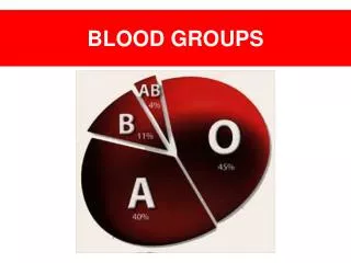
ABO Markers ABO – glycoproteins Which one is most common? Least prevalent?
ABO Markers Preformed antibodies called agglutinins in the plasma is unique to the ABO blood groups. Recall your Practical:
ABO INCOMPATIBILITY • reaction of the immune system that occurs if two different and not compatible blood types are mixed together. • Surface Antigens can act as immune system triggers. • Being exposed to another type of blood can cause a reaction. • Blood types must be matched to avoid an ABO incompatibility reaction. • Important when a patient needs to receive BLOOD (TRANSFUSION) or have an ORGAN TRANSPLANT. • Because type O lacks Ags, type O blood does not cause an immune response. • This is why type O blood cells can be given to patients of any blood type. People with type O blood are called "universal donors." • However, people with type O can only receive type O blood. • Since antibodies are in the liquid part of blood (plasma), both blood and plasma transfusions must be matched to avoid an immune reaction.
ii. THE Rh SYSTEM • Discovered by Landsteiner & Wiener in 1937 • Antigen discovered in the Rhesus monkey • Rh is an Ag on the RBC • a. Rh+ has the Ag (85% of the population) • b. Rh- does NOT have the Rh antigens • c. Rh+ can accept Rh+ or Rh- blood • d. Rh- can accept ONLY Rh- blood
Inheritance of ABO & RH • ABO & RH genes are not linked • ABO & Rh(D) type are inherited independently. • Quiz: Can an A Rh(D)+ mother and a B Rh(D)+ father have an O Rh(D) neg child?
Inheritance of ABO and Rh(D) Mother Group A AO Rh(D) + Dd Father Group B BO Rh(D) + Dd Group A AO Rh(D) pos Dd Group B BO Rh(D) pos Dd Group O OO Rh(D) neg dd Ans: Yes, An A Rh(D)+ mother and a B Rh(D)+ father can have an O Rh(D) neg child.
Rh Factor There are 45 different types of Rh agglutinogens. Common : C, D, and E ~ 85% of Americans are Rh positive, carrying the D antigen. Presence of the Rh agglutinogens on RBCs is indicated as Rh+ As a rule, a person’s ABO and Rh blood groups are reported together, for example, O+, A-, and so on.
Rh Antigens • D antigen is the most common and most immunogenic • Approximately 80-85% Caucasians have D antigen • Individuals lacking this allele are called “Rh-negative” • Only develop antibodies against the D antigen after exposure (transfusion/pregnancy)
Rh antibodies • IgG class of immunoglobulins • Lack capacity to bind complements Hemolysis does not occur after 1st transfusion with Rh factor but it occurs in later transfusions. Why?
Significance of Rh(D) • 80% of Rh(D) negv persons exposed to Rh(D) positive blood will develop anti-D. • Anti-D can also be stimulated by pregnancy with an Rh(D) positive baby • Sensitisation can be prevented by the use of anti-D immunoglobulin, antenatally and post natally • Rh(D) neg females of childbearing potential should never be given Rh(D) positive blood products
Rh AND THE PREGNANT WOMAN • Transplacental passage of D-positive fetal RBC’s into D-negative mother produces anti-D (IgG) • Anti-D IgG traverses the placenta and coats fetal RBC’S leading to extravascular hemolysis • Clinically manifests as hemolytic disease of the fetus and newborn - anemia, hepatosplenomegaly, hydrops fetalis, and death! Background Hydrops fetalis (fetal hydrops) : a serious fetal condition defined as abnormal accumulation of fluid in 2 or more fetal compartments, including ascites, pleural effusion, pericardial effusion, and skin edema.
Rh Incompatibility • Antibodies against the Rh+ factor develop in Rh- person after the first exposure from transfusion. • A clinical problem - if they receive 2nd transfusion of Rh+ blood - Rh Ab will agglutinate with the Rh Ag Rh- mother and Rh+ father can also develop Rh problems with having children. If the mother has a baby that is Rh+, her body will develop antibodies to the Rh antigen so that a second pregnancy with a Rh+ baby will result in the mother’s antibodies attacking the unborn child’s RBCs. Ans: [HDN, or Erythroblastosis fetalis] • Prevention : A shot of RhIg @ RhoGam™ is given shortly after birth to block the development of antibodies. • Symptoms of transfusion reaction: chills and fever, rash, itching, SOB, nausea, nephralgia, hematuria, shock and death.
Erythoblastosis fetalis @ HDN • Disease of fetuses and newborn infants • In transfusions, Rh system is 2nd important to ABO system. • Rh incompatibilitymost common & severe cause of HDN. • Can happen when an Rh-negative woman and an Rh+ man produce an Rh+ baby. The baby has inherited the Rh+ Ag from the Father, and the Mother has developed anti-Rh Ab in response to the Ag. • Most Rh-negative people who receive Rh+ blood will develop anti-D Ab. A later transfusion of Rh-positive blood could result in a severe or fatal transfusion reaction. • These Ab [agglutinins] can diffuse through the placenta into the fetal circulation and cause RBC agglutination. • enter the mother's bloodstream, causing the mother to make anti-D antibodies.
Erythoblastosis fetalis @ HDN…. • Unlike ABO Ab’s, the structure of anti-D Ab makes it possible to cross the placenta and enter the baby's bloodstream. • Specifically, the mother's anti-Rh+ antibodies agglutinate her infant's Rh+ blood. • Symptoms include life threateninganemia, jaundice, fever, swollen tissues from edema, and an enlarged liver and spleen. • Serious cases of this condition are treated by fetal blood replacement.
Preventing Erythroblastosis Foetalis @ HDN First step: Find out the Rh types of the expectant parents. • If mother is Rh-negative and father is Rh-positive, baby is at risk for developing HDN. The next step: Mother's serum is tested to make sure she doesn't already have anti-D Ab from a previous pregnancy or transfusion; similar to blood typing. Finally, the Rh-negative mother is given an injection of Rh Immunoglobulin (RhIg)/ RhoGam™ at 28 weeks of gestation and again after delivery, if the baby is Rh+. The RhIg will attach to any Rh+ cells from the baby in the mother's bloodstream, thus preventing them from triggering anti-D Ab production in the mother. The Rh- woman should also receive RhIg following a miscarriage, abortion, or ectopic pregnancy.
RhoGAM Injection of anti-Rh antibodies given soon after every delivery, miscarriage, abortion-binds Inactivates fetal Rh antigens so mother’s immune system doesn’t respond
Complications in Blood Transfusions Allergic reaction, Air embolism, Citrate intoxication, Hypothermia, Hyperkalemia, Hemosiderosis, Mismatched transfusion.
Transfusions • If mismatched blood given antibodies bind to it and hemolyze cells • Type AB has no AB antibodies so can receive any ABO type blood called Universal recipients • Type O have neither antigen so can donate to any other ABO type called Universal donors • Misleading: because of many other blood groups must be matched also. • Other blood groups: example: • Duffy-negative blood occurs much more frequently in people of African origin • hh antigen system (also known as the Bombay blood group) individuals who can only receive blood safely from other hh donors, because they form antibodies against the H substance
Transfusion Reactions… Transfusion reactions require immediate recognition, laboratory investigation, and clinical management. If a transfusion reaction is suspected during blood administration, the safest practice is to stop the transfusion and keep the intravenous line open with 0.9% sodium chloride (normal saline). Check of the information on the blood unit label and the patient's identification should be performed to ensure that the "right" blood unit was administered to the "right" patient. In most cases, the residual contents of the blood component container should be returned the blood bank, together with a freshly collected blood sample from the patient, and a transfusion reaction investigation should be initiated.
Transfusion Reactions During or within 24 hours of a blood transfusion; Acute transfusion reactions present as adverse signs or symptoms; reactions include fever, chills, pruritus, or urticaria. Other signs include severe shortness of breath, red urine (see pic), high fever, or loss of consciousness [may be the first indication of a more severe potentially fatal reaction].
Agglutination Unagglutinated blood smear Agglutinated blood When different types of blood are mixed within the body, the reaction can be a hemolysis as well as agglutination. Depending on the blood types of the donor and the recipient, mismatching can result in death or no problems at all. Different types of blood are recognized on the molecular level and sometimes rejected by being destroyed and ultimately filtered out by the kidneys in order to expel them from the body along with urine. In the case of a transfusion mistake, worst case scenario would be kidney failure and death. Why? when the kidneys try to filter the blood, they essentially become clogged as they are overwhelmed and cease being effective filters. Additionally, there is a rapid depletion of blood clotting factors which causes bleeding from every body orifice.
Compatible √ Not compatible !!
Symptoms of ABO incompatibility Back pain Fever Blood in urine Jaundice Feeling of ‘impending doom’ Anaphylaxis Signs and tests of ABO incompatibility Bilirubin level is high Complete blood count (CBC) shows damaged RBCs andprobably mild anemia Decreased platelet count; increased fibrin degradation Lab test (perhaps a repeat one) shows incompatibility
Treatment of ABO incompatibility Intravenous fluids Drugs to raise BP if it drops too low Steroids & antihistamines – to treat swelling and allergies Promote diuresis Hemodialysis for disrupted renal function Complications of ABO incompatibility: Immunological effects : Hemolysis, urticaria, noncariac pulmonary edema, anaphylaxis Non-immunological effects:Fever with shock, Kidney failure, congestive heart failure, hypotension, death (if not treated)
Summary: ABO INCOMPATIBILITY Donor blood + Recipient Ab ACTIVATES Complement System Acute circulatory problem Anuria DIC Profuse Bleeding Intravascular hemolysis Hemoglobinemia Hemoglobinuria DEATH One of the most lethal effect of TRANSFUSION: RENAL FAILURE Excess Hb from the hemolised RBCs leaks through the glomerular membranes into the renal tubules. Reabsorbtion of water from the tubules causes Hb conc. to rise; resulting in the HB precipitation and subsequent blockade of the tubules.
DIC- Disseminated Intravascular Coagulation DEFN: a serious disorder in which the proteins that control blood clotting become abnormally active. Causes Normally in injury, certain proteins in the blood become activated and travel to the injury site to help stop bleeding. However, in DIC patients, these proteins become abnormally active; due to inflammation, infection, or cancer. Small blood clots clog up the vessels and cut off blood supply to organs eg. liver, brain, or kidney; ORGAN DAMAGE Over time, the clotting proteins are consumed or "used up" ; the person is then at risk for serious bleeding, even from a minor injury or without injury. This process may also break up healthy red blood cells. DIC can be life-threatening! Risk Factors: Blood transfusion reaction Cancer, esp. leukemia. Pregnancy complications (such as placenta that is left behind after delivery) Recent surgery or anesthesia Sepsis; Severe tissue injury (as in burns and head injury) Symptoms Bleeding, possibly from multiple sites in the body Blood clots Bruising Drop in BP Treatment: Blood clotting factors may be replaced with plasma transfusions. Platelet transfusions can raise the blood count. Heparin, a medication used to prevent clotting, is sometimes used to interrupt clotting events. a.k.a: Consumption coagulopathy • http://www.nlm.nih.gov/medlineplus/ency/article/000573.htm
Cross matching – major and minor • Crossmatching is the final step in pretransfusion testing. • Commonly referred to as compatibility testing, or "Type and Cross." • Donor’s RBCs are mixed with recipient’s serum and examined if whether there is any agglutination or not [MAJOR X-matching]. • Also, admix recipient’s RBCs with donor’s serum [MINOR X-matching]. • Observe: if there is an agglutination or not. • NO AGGLUTINATION: in either case means that the two blood are perfectly COMPATIBLE: Transfusion is allowed. Importance of Cross matching: It is a direct and final check to detect whether there is any mismatching btw the bloods of potential donors and potential recipient.
Cross matching … • Before blood from a donor and the recipient are crossmatched, both are ABO and Rh typed. • Also, antibody screening is done to look for Ab to certain Rh, Duffy, MNS, Kell, Kidd, and P system antigens. • If an antibody to one of these antigens is found, only blood without that antigen will be compatible in a crossmatch. • To begin the crossmatch, blood from a donor with the same ABO and Rh type as the recipient is selected. • In a test tube, serum from the patient is mixed with RBCs from the donor. If agglutination/clumping occurs, the blood is not compatible; if clumping does not occur, the blood is compatible.
Cross matching … • IF TIME IS AVAILABLE for blood typing, RBCs of the recipient type (type specific cells) are given. • During an EMERGENCY, TIME factor may force the initial use of O-RBCs & preferably Rh-negative. • Why? O-blood type, the universal donor has no ABO antigens for a patient's antibodies to attack. • In contrast, AB blood type, the universal recipient, has no ABO antibodies to attack the antigens on transfused RBCs. • In either case, the cross-match is continued, even though the transfusion has begun; as a precaution.
Major and Minor Cross matching of blood Donor’s Blood Patient’s blood Minor Cross matching Plasma MajorCross matching RBCs Patient’s Plasma + Donor’s RBCs = Major Cross-Match Donor’s Plasma + Patient’s RBCs = Minor Cross-Match TASK: Mix and place a drop of plasma or RBC onto slide/test tube Observe for Agglutination or Hemolysis.
What is Apheresis? • The process of apheresis involves removal of whole blood from a patient or donor. Within an instrument that is essentially designed as a centrifuge, the components of whole blood are separated. One of the separated portions is then withdrawn and the remaining components are retransfused into the patient or donor. • The components which are separated and withdrawn include: • Plasma (plasmapheresis) • Platelets (plateletpheresis) • Leukocytes (leukapheresis) • During centrifugation; • blood separates into components • (P = plasma; PRP = platelet rich plasma; WBC = leukocytes; RBC = red blood cells) • The component to be removed can be selected by moving the level of the aspiration device at the right. In this example, plasma is being removed.
The process of apheresis has become essential in providing blood components for therapy. • A volunteer donor will undergo apheresis to supply specific components. The process takes a few hours. • Examples include: • Plateletpheresis: this is the most common means for supplying HLA matched platelets to patients who have become HLA sensitized and require platelets from a single donor whose HLA type matches theirs. • Plasmapheresis: the plasma can be removed to supply blood components such as clotting factors. Donors can give plasma via this mechanism more often than they can donate whole blood. • Leukapheresis: the leukocytes (specifically the granulocytes) can be harvested from a donor to supply granulocytes to help fight infection in patients such as neonates. In some cases of leukemia with very high white blood cell counts, removal of the excess leukocytes may help to prevent complications of thrombosis.