Download
1 / 30
300 likes | 430 Views
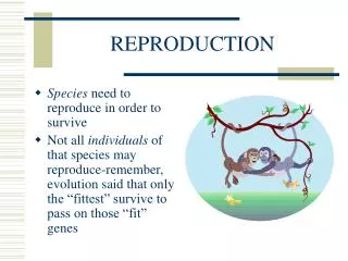
Reproduction. Chris Ellason. Anatomy of Pituitary. Two distinct lobes of different anatomical structure and embryological origin. Anterior pituitary a.k.a. adenohypophysis. Pars distalis Pars tuberalis Posterior pituitary a.k.a. neurohypophysis. Pars nervosa Pars intermedia.

E N D
Reproduction Chris Ellason
Anatomy of Pituitary • Two distinct lobes of different anatomical structure and embryological origin. • Anterior pituitary a.k.a. adenohypophysis. • Pars distalis • Pars tuberalis • Posterior pituitary a.k.a. neurohypophysis. • Pars nervosa • Pars intermedia
Anterior Pituitary • Highly vascular endocrine gland that is derived from an invagination of the roof of the mouth of the developing embryo. • Pars distalis occupies the majority of the mass of the adenohypophysis. Localized regions of this gland produce F.S.H., L.H., L.T.H., S.T.H., T.S.H., A.C.T.H. and M.S.H. (in mammals, but not birds).
Anatomy Cont • The pars tuberalis is a thin sheet of epithelial tissue that serves to separate the pars distalis from the pars nervosa of the neurohypophysis. It serves no known endocrine function. • The pars intermedia is present in birds, reptiles and amphibians and in those species produce melanocyte stimulating hormone (M.S.H.).
Pars Nervosa • The pars nervosa occupies the entire mass of the posterior pituitary in mammals and most of it in birds. • It is made of neurosecretory axons and dendrites and is derived embryologically from the developing hypothalamus. • It serves as a storage and releasing gland for oxytocin and vasopressin from the hypothalamus.
Follicle Stimulating Hormone • Produced and released from pars distalis under GnRH control • Acts on ovary to promote growth and development of young ovarian follicles
Follicular Development • Primary follicle- single layer of cells surrounding an oocyte • Secondary follicle- layers grow and divide to form multiple layer of cells surrounding the oocyte. Zona Pellucida added.
Zona Pellucida • Second membrane that surrounds the ovum • Selectively impermeable to supernumerary sperm preventing multiple fertilizations of the ovum • Important in embryo survival • Loses this ability as the ovum ages in the oviduct
Development cont • Tertiary follicle- Inner cell layer begins secretion of fluid and the formation of an antrum (pocket) • Graafian follicle- Secondary oocyte perched on a pedestal of cells the cumulus oophorus. Fluid filled blister up above the surface of the ovary. Ready to ovulate. Has been secreting estrogens since the late secondary follicle stage
FSH in male • Stimulates growth and development of the seminiferous tubules. These occupy the mass of the testes and produce sperm • Responsible for the stimulation of change from various stages of spermatogensis • Thus in both male and female, FSH acts on a similar function, I.e. gamete development and delivery
Luteinzing Hormone • Produced in the pars distalis and released by action of GnRH from hypothalamus • The LH surge stimulates the apical region of the Graafian follicle to weaken and combined with fluid pressure stimulates ovulation (release) of the ovum from the follicle into the infundibulum
LH cont • Continuing under the influence of LH the ovulation site becomes the corpus hemorrhagicum. The luteal cells increase in number and the corpus luteum is formed over a period of 4 days. By day 8 the CL is at a maximum • The CL is maintained throughout the cycle or pregnancy by the action of LH
LH in the Male • In the male LH is known as ICSH, interstitial cell stimulation hormone. However, they are the same hormone chemically • The interstitial cells or Cells of Leydig are found in the testes interstitial (in between) to the seminiferous tubules. These cells produce the male hormone testosteone
Secretion Differences • During the indifferent stage of embryonic development the presence of androgens in the male embryo imprints the male secretion pattern on the hypothalamus • Without androgens the female embryo takes on the default pattern of a cyclic release of GnRH
Luteotropic Hormone • Proteinaeous gonadotropic hormone produced in the pars distalis of the anterior pituitary • From the hypothalamus there are two different releasing hormones produced that affect the release or non-release of LTH • These are PRH and PIH
LTH cont • Prolactin releasing hormone (PRH) from the hypothalamus stimulates a release of prolactin or LTH which acts on the corpus luteum to stimulate progesterone production and release • As LTH rises in the blood and hits its preset threshold, PIH or prolactin inhibiting hormone is released from the hypothalamus
LTH cont • PIH inhibits the release of LTH from the pars distalis and thus reduces the level of progesterone in the blood • Thus, the two hormones from the hypothalamus moderate or keep the level of progesterone at a constant level in the blood by means of progesterone feeding back to the hypothalamus
LTH cont • As long as progesterone remains in the blood because of LH maintenance of the CL and LTH stimulation of the CL, the levels of progesterone will be maintained at a constant level • As a side effect, LTH inhibits the release of GnRH by the hypothalamus • Remember: LTH high, no GNRH, no FSH, no cycle
LTH cont • LTH is also known as prolactin. This terminology is due to the fact that prolactin is responsible for the alveolar cells of the mammary gland producing milk in mammals • However, after parturition there is no CL on the ovary and thus no progesterone is produced
LTH cont • An alternative control of prolactin is in place for the non-pregnant lactating female • Prolactin is released as long as the mammary gland is empty or only partially full. When the udder is full then PIH is released and prolactin declines. It stays low until the udder has some release of milk
LTH cont • In most mammalian species, the young when first born suckle often thus keeping the udder only partially full. Thus, LTH is always high in the blood to manufacture more milk. Remember • This causes what is termed as postpartum anestrus which will last until the young is weaned or becomes mature enough to gain nourishment from outside sources
Placental Gonadotropins • The placentas of certain mammals produce species specific hormones • The pregnant mare produces PMSG (Pregnant Mare Serum Gonadotropin) • The pregnant women produces HCG (Human Chorionic Gonadotropin)
PMSG • Produced by the developing placenta beginning about day 60 in the mare • Large protein hormone that is recovered from the blood serum • In the mare stimulates secondary corpora lutea. The CL of ovulation regresses early in pregnancy and must be replaced
PMSG • PMSG has FSH like properties. • When used exogenously, in other species it will cause the development of more than normal number of follicles • E.g. Day 18 of cycle give PMSG and boost the number of follicles developed
HCG • Protein hormone produced by the chorion of the developing placenta in the pregnant woman • Has LH like properties and is the basis behind the early pregnancy test that is marketed in drug stores
Estrus Synchronization Manipulation of the estrus cycle to synchronize ovulation and breeding of female groups Generally focus on use of prostaglandin injection protocols to lyse the corpus luteum
PMSG & HCG Application • Day 18 of normal cycle in most domestic females give PMSG. Induce the formation of supernumerary follicles • Day 21 of normal cycle give HCG which will ovulate the extra follicles and cause superovulation • Breed on estrus and collect eggs about day 7 to 9 of pregnancy
Superovulation Manipulation of ovulation to dramatically increase simultaneous release in a single genetically superior individual Generally involves use of follicle stimulating hormone and prostaglandin Multiple ova may be fertilized in vivo or in vitro Ova are manually collected from the dam Recipient females are utilized to implant fertilized ova
Embryo Transfer Transferring embryos from one genetically superior female to other females Embryos may be recovered from a donor female or may be fertilized and grown in vitro Often linked with superovulation Transfer of frozen embryos also common, allowing longer-term storage