Download
1 / 30
320 likes | 453 Views
Delve into the world of nucleic acids, DNA structure, replication, and the flow of genetic information in living systems. Learn about the central dogma of molecular biology and key players in elucidating DNA's structure. Explore DNA replication, nucleotide subunits, Watson-Crick model, and more.

E N D
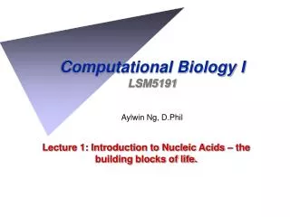
Computational Biology ILSM5191 Aylwin Ng, D.Phil Lecture 1: Introduction to Nucleic Acids – the building blocks of life.
DNA & CHROMOSOMES 2m of DNA, all 3 billion letters in the DNA code, compacted into 46 chromosomes, and packed into a cell 0.0001cm across!
Flow of Information in Living Systems Central Dogma of molecular biology: transcription translation DNA RNA Protein DNA Sequence Implies Structure Implies Function
Transcription mRNA Transport Translation Nascent polypeptide mRNA ribosome Post-transl. modif functional protein
NUCLEIC ACIDS • Deoxyribonucleic acid (DNA) contains the information prescribing the amino acid sequence of proteins. • This information is arranged in units termed genes. • A GENE is the entire nucleic acid sequence that is necessary for the synthesis of a functional polypeptide • Ribonucleic acid (RNA) serves in the cellular machinery that chooses and links amino acids in the correct sequence. • DNA and RNA are polymers of nucleotide subunits
Nucleotide Phosphate group Base Ribose or Deoxyribose (shown here) NUCLEOTIDE SUBUNITS • A nucleotide unit consists of a pentose sugar, a phosphate moiety (containing up to 3 phosphate groups) and a Base. • Subunits are linked together by phosphodiester bond, to form a ‘sugar-phosphate backbone’:
NUCLEOTIDES • All nucleotides have a common structure
BASES • 5 principal bases in nucleic acids: A, G, C, T are present in DNA A, G, C, U are present in RNA
ELUCIDATING THE STRUCTURE OF DNA • James Watson (Cambridge University), • Francis Crick (Cambridge University), • Maurice Wilkins (King’s College London), • Rosalind Franklin (King’s College London) • - succeeded in obtaining superior X-ray diffraction data Nobel Prize (Medicine) in 1962
X-RAY DIFFRACTION • Data showed that DNA has the form of a regular helix • Diameter 20 Å (2 nm) • Making a complete turn every 34 Å (3.4 nm) • i.e. 10 nucleotides per turn
BASE-PAIRING Edwin Chargaff’s results (1952): Base compositions experimentally determined for a variety of organisms
DNA STRUCTURE • Native DNA (B-form) is a double helix of complementary anti-parallel chains. • Double helix is right-handed, with turns running clockwise along helical axis. Hydrogen bonding between complementary base pairs (A-T or G-C) holds the two strands together
DNA REPLICATION • “It has not escaped our notice that the specific pairing we have postulated immediately suggests a possible copying mechanism for the genetic material.” • Watson & Crick, Nature (1953)
DNA REPLICATION • DNA replication is semi-conservative. • IMPLICATION: • the structure of DNA carries information needed to perpetuate its sequence . • Demonstrated by Meselson-Stahl (1958) • Labeled parental DNA with ‘heavy’ density label by growing E. coli in medium containing isotope (e.g. 15N): Light (14N) Hybrid Heavy (15N) parental 1st Gen 2nd Gen
NUCLEIC ACID SYNTHESIS • Both DNA and RNA chains are produced by copying of template DNA strands. • Nucleic acid strands grow in the 5’ 3’ direction. • Energetically unfavorable. Driven by energy available in the triphosphates. • DNA-dependent RNA polymerases can initiate strand growth but DNA polymerases require a primer strand.
E. coli DNA polymerases Main replicating enzyme DNA repair DNA repair & replication
DNA Replication Clip http://academy.d20.co.edu/kadets/lundberg/DNA_animations/DNAreplication.mov
BIDIRECTIONAL REPLICATION • DNA replication proceeds bidirectionally from a given starting site (Origin of Replication), with both strands being copied at each fork. • Common features of Replication Origins (of E. coli, yeast, SV40) • Unique segments containing multiple short repeated sequences, • Short repeated units recognised by multimeric proteins (which assembles DNA polymerases & replication enzymes), • Origin regions contain an AT-rich stretch (less energy req.d to melt A.T base pairs).
BIDIRECTIONAL REPLICATION • Key events prior to the replication process (E. coli): • Binding of DnaA protein at Origin separate (‘melt’) the strands. • DnaC & DnaB bind at Origin. • Then Helicase (DnaB) unwinding of duplex in opposite directions away from Origin. • Unwinding of duplex is an ATP-dependent process. • Single-strand binding (SSB) protein binds to the single-stranded (ss) DNA, preventing it from reforming the duplex state. • Primases (RNA polymerase) bind to DnaB helicase primosome complex • Primases dissociate after synthesizing short primer RNAs (complementary to both strands).
MAMMALIAN DNA POLYMERASES Main replicating enzyme Priming DNA repair Mitochond. DNA replication
REPLICATION IN EUKARYOTES • Very similar to replication in bacteria, differing only in details. • DNA polymerase has primase activity generates RNA primers. • DNA polymerase is the main replicating enzyme. • Eukaryotic DNA polymerases appear to lack 5’ 3’ exonuclease activity needed to remove RNA primer from each Okazaki fragment. • ‘Flap endonuclease’ (FEN1) initiates primer degradation by associating with DNA polymerase .
What happens at Telomeres? Leading strand Chromosome end (Telomere) 3’ 5’ 5’ Parent molecule 3’ 3’ 5’ Lagging strand 3’ 5’ 5’ 3’ 2 Daughter molecules 200bp 200bp 200bp 200bp 200bp 3’ 5’ Missing Okazaki fragment 5’ 3’ 5’ Next Generation (or Grand-Daughter) molecule 3’ 5’ 3’ Molecule has become shorter
The Solution: TELOMERASE • Telomeres can be extended by an independent mechanism. • Catalysed by TELOMERASE. • Enzyme consists of both protein & RNA. • RNA is 450 nucleotides long. • Contains the seq. 5’-CUAACCCUAAC-3’ near its 5’ end. • Underlined seq. is the reverse complement of the human telomere repeat seq. 5’-TTAGGG-3’. • This allows telomerase to extend the 3’end a sufficient amount, • to facilitate priming & synthesis of a new Okazaki fragment by DNA polymerase • generate a double-stranded end.