Download
1 / 42
420 likes | 753 Views
Urinary disorder in pregnancy by: Dr.Rozhan yassin khalil. Structure of the lower urinary tract :. In women the lower urinary and genital tracts develop in close proximity.

E N D
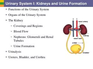
Structure of the lower urinary tract: • In women the lower urinary and genital tracts develop in close proximity. • The bladder is a hollow muscular organ normally situated behind the pubic symphysis and covered superiorly and anteriorly by peritoneum. • The normal adult female urethra is between 3 and 5 cm in length It is a hollow tubular structure joining the bladder to the exterior and is located under the pubic symphysis.
Urinary tract infection: Acute and chronic urinary infections are important and-avoidable sources of ill-health among women. • predisposing factor of urinary tract infection : • 1- short urethra, which is prone to entry of bacteria during intercourse, • 2- poor perineal hygiene and • 3- the occasional inefficient voiding ability of the patient and • 4- unnecessary catheterizations are all contributory factors.
Urinary tract infection in pregnancy: During pregnancy the ureters are dilated and kinked because of: • Increased progesterone levels which relax the smooth muscle. • Mild obstruction of the lower ureters in late pregnancy. This encourages: • Stasis of urine. • Reflux of infected urine to the kidney evoking pyelonephritis.
In pregnancy: -During pregnancy, high progesterone levels elevate the risk of decreased muscle tone of the ureters and bladder, which leads to a greater likelihood of reflux, where urine flows back up the ureters and towards the kidneys. if bacteriuria is present they do have a 30% risk of a kidney infection. Thus if urine testing shows signs of an infection—even in the absence of symptoms,treatment should start
Urinary Tract Infections: • Leading cause of morbidity and health care expenditures in persons of all ages. • An estimated 50 % of women report having had a UTI at some point in their lives.
Types: of UTI • Asymptomatic bacteriuria • Cystitis • Pyelonephritis • Urethritis Asymptbacteriuria, acute cystitis and acute pyelonephritis are common in pregnancy
Asymptomatic bacteriuria: The presence of more than 100,000 bacteria/ml of urine in the absence of symptoms. • Incidence: • About 3% of pregnant women—increases with parity and age. • Significance: • Asymptomatic bacteriuria is associated with a risk of Acute pyelonephritis in pregnancy (30%).
Acute Uncomplicated Cystitis • The microbiology is limited to a few pathogens. • 70%- 85% are caused by Escherichia coli • 5-20%are caused by coagulase-negative Staphylococcus saprophyticus • 5-12% are caused by other Enterobacteriaceae such as Klebsiella and Proteus.
These pathogene transport via: • 1-These gain entry to the urinary tract by a direct extension from the gut. • 2- lymphatic spread. • 3- via the bloodstream or. • 4-transurethrally from the perineum.
• Structural abnormalities in the urinary tract (3–5%). Screening: In early pregnancy, all women should have urine tested for either: • The presence of white cells and nitrites, or; • cultured for bacteria.
Treatment: The most common organisms grown are: • Escherichia coli. • Proteus mirabilis. These are usually sensitive to amoxycillin or nitrofurantoin,cephalexin, Ampicillin . A 5-day course of an antibiotic to which the organism is sensitive should be prescribed.
Treatment: • drugs as • 1-garamicin • 2- ciprofloxacin. • 3- trimethoprim. (sulpha ). • All contraindicated during pregnancy.
Treatment: This will result in a cure in more than 85% of women, but the urine should be recultured one week after treatment. A renal ultrasound and an intravenous (i.v.) urogram should be performed 3 months after delivery to exclude a structural urinary tract abnormality.
Acute pyelonephritis: • Occurs in 1-2% of all pregnant women • Usually follows asymptbacteriuria • Associated with risk to mother and fetus • Common cause of hospitalisation during pregnancy • Maternal effects include fever, toxic shock, renal insufficiency, anaemiaetc • preterm labor and fetal death can occur in severe cases. • Treat for 10-14 days.
Symptomatic infections: Incidence 1–2%; commoner in primigravidae Symptoms: • Dysuria (due to urethritis). • Increased frequency (due to trigonitis). • Backache, loin pains, night sweats and rigors (due to pyelonephritis). • Headache, vomiting and muscle aches (due to pyrexia).
Symptomatic infections: Examination: • The woman is usually pyrexial if the infection has involved the kidneys. In many cases this may be at levels of up to 40.5°C. • If the woman has pyelonephritis she will be tender in the renal angles.
Investigation: A mid-stream specimen of urine (MSU) should be sent for: • Dipstick for nitrites and leucocytes. • Microscopy for white cells. • Culture to determine the organism responsible. • Sensitivity of organisms to antibiotics.
Management: All women who have renal angle tenderness or a pyrexia must be admitted to hospital because of the threat of preterm labour. Management consists of: 1 Bed rest. 2 Ample fluid intake, at least 3 litres a day; if nauseated, give i.v. 3 Start a broad-spectrum i.v. antibiotic such as amoxycillin; may need changing when an organism’s antibiotic sensitivities are known.
Management conti: 4 If the woman has pyelonephritis, do a renal ultrasound when she has recovered from the infection. 5 Keep her in hospital until the renal angle tenderness has disappeared. 6 The antibiotics can be given orally once temperature is normal. A complete 5-day course at least should be given.
Chronic renal disease: Renal changes in normal pregnancy: •1- Renal blood flow increases. •2- Glomerular filtration rate (GFR)increases. •3- Plasma concentrations of urea and creatinine fall in normal pregnancy.
Chronic renal disease: • There is an increase in total body water that exceeds the increase in total body sodium, resulting in a decrease in plasma osmolality. • There is a 25% fall in serum uric acid concentrations during the first two trimesters but this returns to pre-pregnant levels by the third trimester. Watch if using urate to monitor pre-eclampsia.
Prognosis: The outcome of the pregnancy is worse if: 1- The woman was hypertensive before pregnancy. 2- The woman hadproteinuriabefore pregnancy started. 3 -There is active progression of renal disease or it is associated with other medical conditions.
Prognosis Pregnancy probably has no long-term adverse effects on renal disease. Fetal prognosis: • Normotensive women with chronic renal disease have 2–3 times greater risk of developing pre-eclampsia. In the absence of pre-eclampticperinatal mortality is not increased. If PE develops, the risk of fetal death is directly related to the gestation at delivery.
• Fetal prognosis: Women with more severe renal disease have a high incidence of both PE and impaired fetal growth. Among women with pre-existing hypertension and proteinuria, the perinatal mortality rates approach 30%. Cause of death is from preterm delivery and complications associated with small for gestational age (SGA).
Acute renal failure in pregnancy: • This may be: • 1- Tubular necrosis: this is largely reversable. • 2- Cortical necrosis: this is usually irreversable • and these patients go on to need long-term dialysis • or transplantation.
Acute renal failure presentation 1 Oliguria: <500ml/day (20ml/hour), the minimum volume to remove catabolites. 2 Anuria: the absence of urine. Aetiology in obstetrics: 1 Hypovolaemia. • Severe pre-eclampsia. • Placental abruption. • Postpartum haemorrhage (PPH). • Hyperemesisgravidarum. • Miscarriage.
Aetiology in obstetrics: 2- Gram-negative shock. This may result from: • Pyelonephritis. • Chorioamnionitis. • Puerperal infections. • Septic miscarriage. The usual organism is E. coli, but it may be Clostridium. 3- Nephrotoxins: In modern obstetric practice : These are rare. Illegal abortions may result in infection followed by haemolysis and renal failure.
Aetiology in obstetrics: 4 Acute renal failure associated with acute fatty liver of pregnancy:Rare; usually fatal. 5 Vomiting in late pregnancy associated with jaundice: The disease occurs in many systems and renal failure, pancreatitis and colitis may occur.
Management: Three consecutive phases: 1- Oliguria: lasts from a few days to a few weeks. Complete anuria is rare in acute tubular necrosis and usually suggests acute cortical necrosis or obstruction. 2 Polyuria: markedly increased urine production that may last up to 2 weeks. The urine is dilute and metabolic waste products are poorly eliminated. Plasma urea and creatinine may continue to rise for several days following the increase in urinary output. Profound fluid and electrolyte losses can occur in this phase.
3- Recovery: urinary volumes decrease towards normal and renal function improves. General management: 1-Determine the cause. 2-Insert a urinary catheter and maintain accurate fluid balance charts.
3- Insert a central venous pressure line and measure the pressure. In pregnancy, central venous pressure should range from +4 to +10cmH2O. If this is low it suggests the cause of the renal failure is due to hypovolaemia and therefore the volume should be restored with up to 2 litresof normal saline followed by a plasma expander. Response of over 30ml of urine in 1 hour should be seen within 1 hour of the fluid load.
4- Send baseline investigations: including urea and electrolytes, liver function tests, serum amylase, plasma proteins, coagulation studies, and if required, perform an arterial blood sample for acid–base balance. • If the patient is not hypovolaemic or does not respond to the fluid load, then the alternatives are as follows: (a) Conservative management. Give i.v. the volume of patient fluid output plus 500ml/day. Monitor electrolytes and start dialysis if or when the patient is uraemic or hypercatabolic.
(b) Intensive management using vasodilators and inotropes (renal dose dopamine). This requires the insertion of a pulmonary artery catheter and the monitoring of cardiac output. Vasodilators and inotropes are given and the patient is fluid-loaded. • Involve intensive care physicians and the renal physician at an early stage.
Pregnancy in patients with chronic kidney disease: • Proteinuria increases in ~ ½ of the patients • Hypertension develops or worsens in ~ ¼ of the patients • Significant worsening of edema can occur during pregnancy in women with nephrotic syndrome • Β-HCG can be increased in patients with ESRD, so confirm pregnancy with an ultrasound
Pregnancy in patients on dialysis • Conception occurs in 0.3-1.5% of women of childbearing age per year (disrupted gonadal function) • Live births occur in 40-50% • Prematurity occurs in most (average age at delivery is ~ 30.5 weeks) • Increased risk for severe hypertension. • More intensive dialysis recommended (5-7x/wk to keep BUN under 45-50); more frequent, lower volume exchanges if on peritonial dialysis. • Avoid hemodynamic instability & monitor the fetus during treatment
Pregnancy in renal transplant patients: outcomes • Fertility returns! • > 90% success after 1st trimester; slight increase in spontaneous abortion • IUGR a/o premature delivery in up to 20% & 50%, respectively . • 14% spontaneous abortion, high prevalence of hypertension, increased preeclampsia . • Developmental delays related to prematurity • Fewer complications & birth abnormalities than dialysis patients
Transplant medications:steroids: • Associations noted between prednisone & a variety of birth defects (but mainly at doses > 20 mg/d) • Retrospective data suggest an increased risk of cleft palate with glucocorticoids • Possible increased risk of PROM & IUGR • Glucocorticoids are excreted in breast milk (small amounts), but considered ok if needed by mother
Transplant medications: cyclosporine: • Can induce/worsen hypertension • Drug levels may fall during pregnancy • Premature labor and infants that are small for gestational age have been reported .
OK to biopsy?? • Data for native kidneys • Can be done safely in women with well-controlled blood pressure • Biopsy after 32 weeks is not recommended (? if applies to transplant patients?)