Download
1 / 4
40 likes | 256 Views
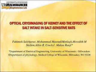
OPTICAL CRYOIMAGING OF KIDNEY AND THE EFFECT OF SALT INTAKE IN SALT-SENSITIVE RATS. Fahimeh Salehpour , Mohammad MasoudiMotlagh,Meredith M Skelton,Allen R. Cowley ǂ , Mahsa Ranji * *Department of Electrical Engineering, University of Wisconsin - Milwaukee

E N D
OPTICAL CRYOIMAGING OF KIDNEY AND THE EFFECT OF SALT INTAKE IN SALT-SENSITIVE RATS FahimehSalehpour, Mohammad MasoudiMotlagh,Meredith M Skelton,Allen R. Cowleyǂ, MahsaRanji* *Department of Electrical Engineering, University of Wisconsin - Milwaukee ǂDepartment of physiology, Medical College of Wisconsin, Milwaukee, WI 53226
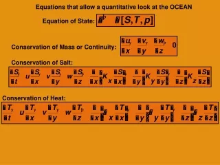
OBJECTIVEAND INTRODUCTION • Hypertension is the biggest risk factor for the development and progression of stroke, heart failure, and end-stage renal disease. • Excess dietary salt consumption plays a causal role in the development of salt-sensitive hypertension and renal oxidative stress and injury. • In this study we used fluorescence imaging in cryogenic temperature to evaluate whether the increased consumption of salt contributes to excess oxidative stress and salt-sensitive hypertension.
10 6 8 7 4 9 3 1 METHODOLOGY 5 A/D Digital I/O Board Frame Grabber • Lens • Stepper Motor • CCD Camera • Tissue Block • Microtome Blade • Excitation Filter Wheel • Emission Filter Wheel • Ultra Low Freezer • Excitation Source • Power Supply 2 • NADH and FADautofluorescent images from each group of kidneys, were processed to extract NADH RR values using MATLAB. • Composite images were created using all image slices from cryoimager for each tissue, for both NADH and FAD signals. • The image stacks were analyzed in MATLAB to measure the 3-D max projection NADH RR of tissue and their histograms.
RESULTS AND CONCLUSION Histograms Salt-sensitive Salt-resistance Mean = 106.55 Std = 23.973 NADH NADH Mean = 135.933 Std = 50.45 28% FAD Mean = 1.993 Std= 0.639 NADH Redox NADH RR Mean = 3.252 Std= 1.326 63%